Download
1 / 19
190 likes | 197 Views
Learn about the female reproductive system, including the ovaries, uterine tubes, uterus, vagina, and external genitalia. Explore the stages of follicular development and the histology of a Graafian follicle. Understand the process of oogenesis and the histology of the uterus. Discover the blood supply to the uterus and the anatomy of the vulva. Gain insights into the female reproductive cycle, including hormonal regulation and the phases of the menstrual cycle.

E N D
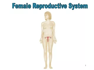
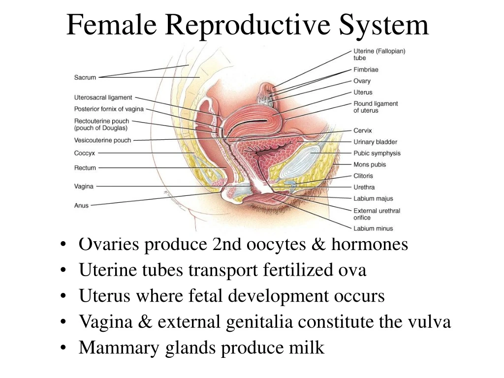
Female Reproductive System • Ovaries produce 2nd oocytes & hormones • Uterine tubes transport fertilized ova • Uterus where fetal development occurs • Vagina & external genitalia constitute the vulva • Mammary glands produce milk
Follicular Stages • Stages of follicular development • primordial • primary • secondary • graafian • ovulation • Corpus luteum is ovulation wound • fills in with hormone secreting cells • Corpus albicans is white scar left after corpus luteum is not needed
Histology of a Graafian Follicle • Zona pellucida -- clear area between oocyte & granulosa cells • Corona radiata is granulosa cells attached to zona pellucida--still attached to oocyte at ovulation • Antrum formed by granulosa cells secreting fluid • By this time, the oocyte has reached the metaphase of meiosis II stage and stopped developing -- first polar body has been discarded
Life History of Oogonia • Germ cells from yolk sac migrate to ovary & become oogonia • As a fetus, oogonia divide to produce millions by mitosis but most degenerate (atresia) • Some develop into primary oocytes & stop in prophase stage of meiosis I • 200,000 to 2 million present at birth • 40,000 remain at puberty but only 400 mature during a woman’s life • Each month, hormones cause meiosis I to resume in several follicles so that meiosis II is reached by ovulation • Penetration by the sperm causes the final stages of meiosis to occur
Histology of the Uterus • Endometrium • simple columnar epithelium • stroma of connective tissue and endometrial glands • stratum functionalis • shed during menstruation • stratum basalis • replaces stratum functionalis each month • Myometrium • 3 layers of smooth muscle • Perimetrium • visceral peritoneum
Blood Supply to the Uterus • Uterine arteries branch as arcuate arteries and radial arteries that supply the myometrium • Straight & spiral branches penetrate to the endometrium • spiral arteries supply the stratum functionalis • their constriction due to hormonal changes starts menstrual cycle
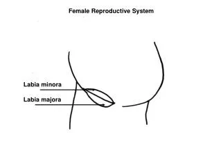
Vulva (pudendum) • Mons pubis -- fatty pad over the pubic symphysis • Labia majora & minora -- folds of skin encircling vestibule where find urethral and vaginal openings • Clitoris -- small mass of erectile tissue • Bulb of vestibule -- masses of erectile tissue just deep to the labia on either side of the vaginal orifice
Female Reproductive Cycle • Controlled by monthly hormone cycle of anterior pituitary, hypothalamus & ovary • Monthly cycle of changes in ovary and uterus • Ovarian cycle • changes in ovary during & after maturation of oocyte • Uterine cycle • preparation of uterus to receive fertilized ovum • if implantation does not occur, the stratum functionalis is shed during menstruation
Hormonal Regulation of Reproductive Cycle • GnRH secreted by the hypothalamus controls the female reproductive cycle • stimulates anterior pituitary to secrete FSH & LH • FSH initiates growth of follicles that secrete estrogen • estrogen maintains reproductive organs • LH stimulates ovulation & promotes formation of the corpus luteum which secretes estrogens, progesterone, relaxin & inhibin • progesterone prepares uterus for implantation and the mammary glands for milk secretion • relaxin facilitates implantation in the relaxed uterus • inhibin inhibits the secretion of FSH
Menstrual Phase • Menstruation lasts for 5 days • First day is considered beginning of 28 day cycle • In ovary • 20 follicles that began to develop 6 days before are now beginning to secrete estrogen • fluid is filling the antrum from granulosa cells • In uterus • declining levels of progesterone caused spiral arteries to constrict -- glandular tissue dies • stratum functionalis layer is sloughed off along with 50 to 150 ml of blood
Preovulatory Phase • Lasts from day 6 to 13 (most variable timeline) • In the ovary (follicular phase) • follicular secretion of estrogen & inhibin has slowed the secretion of FSH • dominant follicles survives to day 6 • by day 14, graafian follicle has enlarged & bulges at surface • increasing estrogen levels trigger the secretion of LH • In the uterus (proliferative phase) • increasing estrogen levels have repaired & thickened the stratum functionalis to 4-10 mm in thickness
Ovulation • Rupture of follicle & release of 2nd oocyte on day 14 • Cause • increasing levels of estrogen stimulate release of GnRH which stimulates anterior pituitary to release more LH • Corpus hemorrhagicum results
Signs of Ovulation • Increase in basal body temperature • Changes in cervical mucus • Cervix softens • Mittelschmerz---pain
Postovulatory Phase • Most constant timeline = lasts 14 days • In the ovary (luteal phase) • if fertilization did not occur, corpus albicans is formed • as hormone levels drop, secretion of GnRH, FSH & LH rise • if fertilization did occur, developing embryo secretes human chorionic gonadotropin (hCG) which maintains health of corpus luteum & its hormone secretions • In the uterus (secretory phase) • hormones from corpus luteum promote thickening of endometrium to 12-18 mm • formation of more endometrial glands & vascularization • if no fertilization occurs, menstrual phase will begin