Download
1 / 29
290 likes | 303 Views
General Concepts of Brain Organization with Relevance to Clinical Neurology Jeanette J. Norden, Ph.D. Professor Emerita Vanderbilt University School of Medicine. CNS: Brain & spinal cord. PNS: Any nervous elements outside of the brain & spinal cord.

E N D
General Conceptsof Brain Organization with Relevance to Clinical NeurologyJeanette J. Norden, Ph.D.Professor EmeritaVanderbilt University School of Medicine
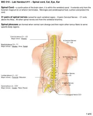
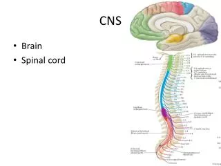
CNS: Brain & spinal cord PNS: Any nervous elements outside of the brain & spinal cord The CNS (brain and spinal cord) is completely surrounded by bone - and is thus within a “closed” compartment
The CNS Develops from a Neural “Tube” The hemispheres will develop over the rostral (front) end The cerebellum will develop over this opening The caudal end will become the spinal cord This caudalmost opening will close off normally at ~4wks
Neurons and Glial Cells are the Major Cell Types in the CNS The Neural Tube will give rise to neurons and other cells of the CNS Oliogdendrocytes Specialized for receiving and transmitting information Myelinate axons Line ventricles Diverse functions
Neurons are Generated (born) Pre-natally • The vast majority of neurons are generated during the first “critical period” of brain development (between 12-20 wks of gestation); during this time, some 200,000 neurons a minute are being “born” • Neurons are “post-mitotic” meaning that they do not divide after they are born • At birth, the brain contains ~200 billion neurons; >50% of these will die post-natally (after birth) so that the adult brain has ~85-100 billion neurons • With rare exception, no new neurons are generated after birth; after birth the brain gets bigger because of addition of other cell types, myelination, etc.
Zika Infects and Kills Neuron Progenitors – resulting in Microcephaly (small head) and Microencephaly (small brain)
NEURONS ARE POLARIZED CELLS SPECIALIZED FOR RECEIVING AND TRANSMITTING “INFORMATION” Neurons communicate electro-chemically + + Myelin “speeds” nerve conduction - Oligodendrocytes These axon terminals will “synapse” with the dendrites/cell body of the next neuron 100 billion neurons interconnected by 100 trillion synapses
NEURONS COMMUNICATE WITH EACH OTHER AT SYNAPSES Neurons communicate “electro-chemically” at synapses Synapses are formed between the axon of one neuron and the dendrites of the next neuron Glutamate (+) and GABA (-) are the major neurotransmitters in the CNS; neuromodulators like dopamine, serotonin “modulate” the activity of neurons that utilize glutamate and GABA Axon terminal of one Neuron (pre-synaptic) SYNAPSE Dendrite of next Neuron (post-synaptic)
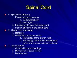
The CNS consists of Functional Groups of Neurons • Groups of neuronal cell bodies and dendrites make up individual structures in the CNS referred to as “nuclei” or “areas”; each of these nuclei/areas has a specific function Lateral view Medial view
Functional Groups of Neurons are Interconnected via Pathways or Axons • Different nuclei/areas are connected to other nuclei/areas by pathways (axons of neurons) to form functional “systems” and interconnected “networks”
Both Anatomy and Chemistry are Important for Normal Functioning of the Brain • For example: groups of neurons called raphe nuclei, which use serotonin as a neurotransmitter, project to other nuclei and areas which are involved in “mood”; thus, mood can be influenced by drugs which affect levels of serotonin; drugs like Prozac elevate mood in some individuals who are depressed by increasing the levels of serotonin available at these synapses • Thus, in development, neurons have to form nuclei (migrate from the area in the neural tube where they are born), interconnect with other nuclei to form functional networks, and develop appropriate chemical signatures for communication
The Adult CNS has 5 Subdivisions that Develop from 3 Areas of the Neural Tube Note that part of the eye is an outgrowth of the brain – and is thus, CNS tissue Embryo Adult
Each Subdivision of the Adult Brain has many Nuclei/Areas with Different & Specific Functions TELENCEPHALON “Higher-order functions DIENCEPHALON (thalamus & hypothalamus) MESENCEPHALON (midbrain) Reflexes METENCEPHALON (pons & cerebellum) MYLENCEPHALON (medulla) Vital functions
OTHER GENERAL CONCEPTS IMPORTANT FOR UNDERSTANDING THE CNS AND THE CONSEQUENCE OF CNS DAMAGE • Each hemisphere processes information from the opposite side of the body and world and controls the movement of the opposite side of the body; some functions show hemisphere dominance (for example, language) • Decussation (crossing) of pathways (for example, neurons in motor cortex project axons to the opposite side of the spinal cord; thus, the motor cortex on the RIGHT controls movement of the LEFT side of the body); different pathways cross in different areas of the brain, and to different degrees • Ipsilateral (same side); contralateral (opposite side); bilateral (both sides)
OTHER GENERAL CONCEPTS IMPORTANT FOR UNDERSTANDING THE CNS AND THE CONSEQUENCE OF CNS DAMAGE • “Lesions” of the CNS; any damage to neurons or axons; can also be “space-occupying” (tumors, swelling, hemorrhage, etc.) • Most lesions are unilateral, meaning that they occur on one side; changes in function can be ipsilateral, contralateral, or bilateral, depending on the specific areas/pathways affected • Consequence of CNS lesions: loss or change in function – possibly permanently; the CNS is VERY unforgiving of damage
ANATOMY IS THE KEY TO DIAGNOSIS • In order to evaluate Neurological illness, the physician will observe the patient, listen to the patient, and examine the patient • The physician will look at the constellation of signs (what is found on examination) and symptoms (what the patient complains of) • The first step in the clinical evaluation is the anatomical diagnosis (Where is the lesion?; Single site [and specific location] or multiple sites [disseminated disorder]?) • The next step in the evaluation is the etiologic diagnosis (What is the lesion?)
CLINICAL CASE • A 23 yo Caucasian female college student (Becky) comes to student health complaining of problems “seeing” with her right eye • During the history, she reveals that about 6 mos. ago, she had an “episode” where she felt that something was wrong with her legs; she thinks that she had visual problems at this time as well, but that it resolved as did the problem with her legs
CLINICAL CASE • On examination, Becky shows • Optic disc (area where axons of neurons leave the eye) swellingon the right; when the light reflex (reflex that shuts down the pupil in response to light) is tested, it is sluggish on the right compared to the left • Paresis (weakness) of both lower limbs (legs); increased deep tendon reflexes bilaterally; Babinski sign bilaterally (indicates involvement of 1st order neurons – called upper motor neurons – bilaterally) • All other aspects of the neurological exam are within normal limits
Information Critical to this Clinical Case • Because the neural part of the eye is an outgrowth of the brain, the physician can examine the eye and gain information about the integrity of the CNS; the “neural” part of the eye (called the “neural retina”) contains neurons that project their axons into the brain so that the image projected onto the back of the eye can be interpreted (you “see” with your brain, not your eyes) • The eye is the only area of the body in which living blood vessels and a part of the CNS can be viewed directly
“SWELLING” OF THE OPTIC DISC and SLUGGISH PUPILLARY RESPONSE INDICATE INVOLVEMENT OF THE OPTIC NERVE (the nerve that connects the eye with the brain) Optic disc swelling due to optic neuritis (inflammation of the optic nerve) Normal, healthy fundus (back of the eye)
Information Critical to this Clinical Case • Spastic weakness of the legs with a Babinski (a reflex stimulated in the foot) can occur only from damage to neurons located in the area of the cortex that initiates movement of the legs – or to the axons of these neurons that project to the spinal cord; in Becky, these neurons have been involved bilaterally Motor Cortex (area of leg representation) Note that Becky has >1 lesion in different areas of the CNS
DIAGNOSIS IN CLINICAL NEUROLOGY • ANATOMIC DIAGNOSIS: Where is the lesion? • Right optic nerve • Upper motor neurons/axons of the corticospinal tract bilaterally (First order neurons in the motor pathway that project to the spinal cord); lesions could be in brain or spinal cord (before the synapse onto spinal cord neurons that go out to the muscle) • No single site lesion could account for both the optic nerve involvement and the involvement bilaterally of the upper motor neurons/axons; thus, Becky has a “disseminated” or “multi-site” disorder • ETIOLOGIC DIAGNOSIS: What is the lesion? • Most likely: MULTIPLE SCLEROSIS
MULTIPLE SCLEROSIS • Achronic inflammatory demyelinating disease of the CNS; over time will produce multiple “scar” (sclerotic) areas or “plaques” • Autoantibodies attack oliodendrocytes and the myelin sheath of CNS axons; neurons will also be affected; what signs/symptoms the patient has depends on what specific pathways are affected (nearly all pathways in the human brain are myelinated – thus almost any pathway could be involved) When myelin is lost, neurons will not conduct normally; once the myelin sheath is lost, the neuron will eventually die
MULTIPLE SCLEROSIS • Because most pathways in the human CNS are myelinated, MS can involve different pathways in different patients; while patients may show very individual patterns of demyelination (and therefore different signs/symptoms), there are some sites that appear to be more commonly affected; for example, the optic nerve is commonly involved, as is the deep white matter (axons) of the hemispheres PLAQUES Plaques in deep white matter (MRI imaging (left); brain specimen at autopsy (right) Demyelinated optic nerve
MULTIPLE SCLEROSIS • The disorder is characterized by periods of exacerbation (↑ inflammation) and remission (↓ inflammation); thus, it is a disorder which is separated in time and space (the latter referring to multiple sites in the CNS) • MS may be relatively benign – or rapidly progressive • Occurs females > males; primarily affects young adults – thus, an individual could live their entire adult life with this disorder • Interventions include drugs which decrease inflammation; physical therapy and exercise
TAKE-HOME MESSAGE • Any function can be lost with CNS lesions • Physicians use their knowledge of CNS anatomy to pinpoint what specific areas of the brain or spinal cord have been affected; imaging and other types of tests are used to help determine what the lesion is – and to confirm the location of the lesion(s) • Even signs/symptoms which appear to “resolve” or get better should be reported to your primary care physician