Download
1 / 35
450 likes | 710 Views
CNS. Brain Spinal cord. Brain. Receives sensory input from the spinal cord as well as from its own nerves (e.g., olfactory and optic nerves) Devotes most of its volume to processing its various sensory inputs and initiating appropriate and coordinated motor outputs. Spinal cord.

E N D
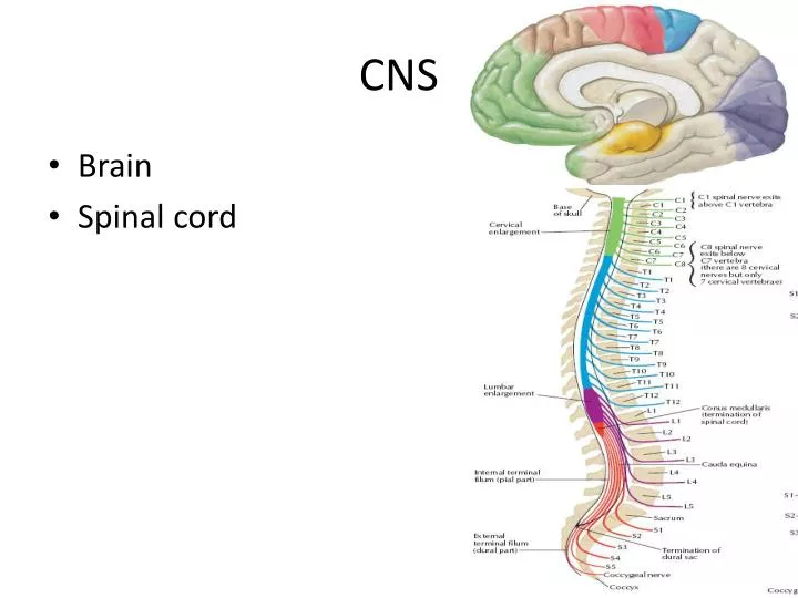
CNS • Brain • Spinal cord
Brain • Receives sensory input from the spinal cord as well as from its own nerves (e.g., olfactory and optic nerves) • Devotes most of its volume to processing its various sensory inputs and initiating appropriate and coordinated motor outputs.
Spinal cord • Conducts sensory informationfrom the peripheral nervous system(both somatic and autonomic) to the brain • Conducts motor informationfrom the brain to our various effectors • Skeletal muscle • Cardiac muscle • Smooth muscle • Glands • Minor reflex center
White and grey matter • Both the spinal cord and the brain consist of white matter and grey matter • White matter= bundles of axons each coated with a sheath of myelin • gray matter = masses of the cell bodies and dendrites —with synapses. • In the spinal cord, the white matter is at the surface, the gray matter inside. • Reverse in the brain
Meninges • Both the spinal cord and brain are covered in three continuous sheets of connective tissue- the meninges. • From outside in • dura mater— pressed against the bony surface of the interior of the vertebrae and the cranium • the arachnoid • the pia mater
Blood-brain barrier • A system of tight junctions between the endothelial cells of the capillaries • Prevents many therapeutic drugs from reaching the brain
Cerebrospinal fluid (CSF), • A secretion of the choroid plexus. • CSF flows uninterrupted throughout the central nervous system • through the central cerebrospinal canalof the spinal cord and • through an interconnected system of four ventricles in the brain.
31pairs of spinal nervesarise along the spinal cord. • Mixed nervesbecause each contain both sensory and motor axons. • Within the spinal column, all the sensory axonspass into the dorsal root ganglionwhere their cell bodies are located and then on into the spinal cord itself. • All the motor axonspass into the ventral rootsbefore uniting with the sensory axons to form the mixed nerves.
Functional areas 1. Motor area (Area No. 4). Precentral gyrus Stimulation of this area results in movements in the opposite half of the body. The body is represented upside down in this area. 2. Sensory area (Area No. 3, 2, 1). Post central gyrus As in the motor area, the body is represented upside down in the sensory area.
3. Premotor area: Located anterior to motor area 6,8, Occupies posterior part of superior, middle & inferior frontal gyri Also called Psychomotor area Pattern of movement are remembered here 4. Motor speech area (of Broca) 45 and 44. Lies in the inferior frontal gyrus (in the left hemisphere in the right handed person and vise versa). Injury to this area results in inability to speak (aphasia).
5. Visual area (Area 17, 18, 19) located in the occipital lobe, mainly in the medial surface, but extends to the superolateral surface 6. Auditory area 41 and 42 (for hearing) is located in the temporal lobe, that is on the superior temporal gyrus
Spinal cord functions • It connects a large part of the peripheral nervous system to the brain. • Information (nerve impulses) reaching the spinal cord through sensory neurons are transmitted up into the brain. Signals arising in the motor areas of the brain travel back down the cord and leave in the motor neurons. • Acts as a minor coordinating center responsible for simple reflexes like the withdrawal reflex. • The interneuron carrying impulses to and from specific receptors and effectors are grouped together in spinal tracts.
The corticospinal tract • The corticospinal tract originates in the cerebral cortex where voluntary motor control is localized. • There are two branches • The lateral: The lateral crosses in the medulla in an area known due to its appearance as the pyramids • These fibers are concerned with control of bilateral postural movements by the supplementary motor cortex. • The anterior: The anterior does not cross
The corticospinal tract • These fibers carried in the corticospinal tract are called "upper motor neurons" and they synapse with "lower" motor neurons in the cord which lead to the skeletal muscles.
Extrapyramidal tracts • Denote all those portions of the brain and brain stem that contribute to motor control but are not part of the direct corticospinal-pyramidal system.
Extrapyramidal tracts • These include pathways through: • the basal ganglia, • the reticular formation of the brain stem, • the vestibular nuclei, and • the red nuclei
Extrapyramidal tracts • The “indirect” or extrapyramidal tracts (outside the pyramids) carry impulses associated with balance and muscle tone to muscles on the opposite side of the body.
Extrapyramidal tracts • Rubrospinal, • Gross motor movements • vestibulospinal, • muscle tone and balance.
Upper motor neuron • Start from cerebral cortex • Ends in the anterior horn cell in spinal cord
Lower motor neuron • Starts from the anterior horn cell in the spinal cord • Ends on the NMJ
Summary Answer the following Questions What are the arteries supplying brain? Summarize in 15 sentences whatever you can remember from previous lecture. Name some pyramidal tracts and their pathways.