Download
1 / 20
2.22k likes | 21.45k Views
Spinal Cord. A. Spinal cord anatomy 1. Protection and coverings a. Vertebral column b. Meninges 2. External anatomy of the spinal cord 3. Internal anatomy of the spinal cord B. Spinal cord physiology 1. Reflexes

E N D
Spinal Cord • A. Spinal cord anatomy 1. Protection and coverings a. Vertebral column b. Meninges 2. External anatomy of the spinal cord 3. Internal anatomy of the spinal cord • B. Spinal cord physiology 1. Reflexes 2. Reflex arc and homeostasis a. Physiology of the stretch reflex b. Physiology of the flexor (withdrawal) reflex and crossed extensor reflexes • C. Spinal nerves 1. Composition and coverings 2. Distribution of spinal nerves 3. Dermatomes
The Spinal Cord • 1. is continuous with brain • 2. mediates spinal reflexes • 3. is site for integration • 4. provides the pathways
Protection and Coverings • 1. vertebral canal • 2. meninges • 3. cerebrospinal fluid
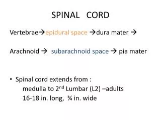
Meninges and Spaces • 1. epidural space • 2. dura mater • 3. subdural space • 4. arachnoid membrane • 5. subarachnoid space • 6. pia mater -- denticulate ligaments
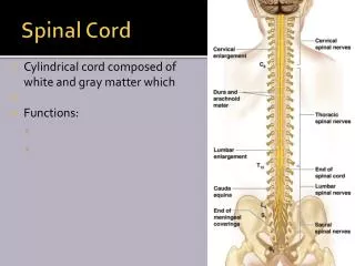
External Anatomy • 1. cylindrical • 2. flattened A-P • 3. foramen magnum to L2 • 4. differential growth • 5. cervical enlargement • 6. lumbar enlargement • 7. conus medullaris • 8. filum terminale • 9. cauda equina • 10. functional segments
Internal Anatomy • 1. gray matter • 2. white matter • 3. gray commissure • 4. central canal • 1. gray matter • 2. white matter • 3. gray commissure • 4. central canal
Gray Matter • 1. nuclei • 2. horns a. dorsal -- sensory b. ventral -- motor c. lateral -- autonomic
Spinal Nerve Roots • 1. dorsal root (axons of sensory neurons) -- dorsal root ganglion (cell bodies of sensory neurons) • 2. ventral root (axons of motor neurons) Dorsal roots Dorsal root ganglion Ventral roots
White Matter • 1. columns a. anterior b. posterior c. lateral • 2. tracts a. ascending b. descending Posterior columns Lateral columns Lateral columns Anterior columns
The Spinal Cord Has Two Essential Functions • 1. convey impulses between the periphery and the brain • 2. provide integrating centers for spinal reflexes
Reflexes are… • 1. inborn • 2. unlearned • 3. unconscious
Somatic Reflexes Versus Visceral Reflexes • Somatic reflexes involve the somatic nervous system. • Visceral reflexes involve the autonomic nervous system.
Reflex Arc 3 2 4 5 4 5 2 1 3 • 1. receptor • 2. sensory neuron • 3. integration center • 4. motor neuron • 5. effector sensory receptor sensory receptor center of integration with association neuron center of integration with association neuron sensory (afferent) neuron sensory (afferent) neuron motor (efferent) neuron motor (efferent) neuron effector effector
THE REFLEX ARC AS A FEEDBACK SYSTEM CONTROLLED CONDITION A stimulus or stress disrupts membrane homeostasis by altering some controlled condition RETURN TO HOMEOSTASIS The action of the effector returns the body process to within its normal homeostatic range RECEPTOR The receptors in a reflex are sensory neurons associated with a receptor device (transducer) and which relay nerve impulses to a central control center EFFECTORS The motor neurons initiate some response by an effector (muscle or gland) to counteract the stimulus that originally disrupted homeostasis CONTROL CENTER The control center is an integrating center of neurons in the CNS. It relays the information to motor neurons
Stretch Reflex • 1. monosynaptic • 2. muscle spindle • 3. muscle tone • 4. ipsilateral • 5. reciprocal innervation
The Flexor and Crossed Extensor Reflex Intersegmental Polysynaptic Ipsilateral Pain receptor Role of association neurons Reciprocal innervation Intersegmental Polysynaptic Contralateral Pain receptor Role of association neurons Reciprocal innervation excitatory neurons inhibitory neurons