Download
1 / 42
430 likes | 699 Views
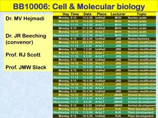
BB10006: Cell & Molecular biology. Dr. MV Hejmadi Dr. JR Beeching (convenor) Prof. RJ Scott Prof. JMW Slack. Dr. Momna Hejmadi (bssmvh@bath.ac.uk). Structure and function of nucleic acids Books (any of these) :

E N D
BB10006: Cell & Molecular biology Dr. MV Hejmadi Dr. JR Beeching (convenor) Prof. RJ Scott Prof. JMW Slack
Dr. Momna Hejmadi (bssmvh@bath.ac.uk) Structure and function of nucleic acids Books (any of these): Any bioscience textbook will do but my favourites are Biochemistry (3e) by D Voet & J Voet Molecular biology of the cell(4th ed)byAlberts et al Essential Cell Biology by Alberts et al Key websites • http://www.dnaftb.org/dnaftb/ • http://www.dnai.org/lesson/go/2166/1994 • http://molvis.sdsc.edu/dna/index.htm
Outline of my lectures Lecture 1. Nucleic acids – an introduction Lecture 2. Properties and functions of nucleic acids Lecture 3. DNA replication Lectures4-6. Transcription and translation Access to web lectures at http://www.bath.ac.uk/bio-sci/hejmadi/teaching%202005-06.htm
Lecture 1 - Outline • How investigators pinpointed DNA as the genetic material • The elegant Watson-Crick model of DNA structure • Forms of DNA (A, B, Z) References: History, structureand forms of DNA http://www.dnai.org/lesson/go/2166
1869 Timeline F Miescher - nucleic acids 1928 F. Griffith - Transforming principle http://www.dnai.org/lesson/go/2166/1994
Discovery of transforming principle • 1928 – Frederick Griffith – experiments with smooth (S) virulent strain Streptococcus pneumoniae and rough (R) nonvirulent strain
What is this transforming principle? • Bacterial transformation demonstrates transfer of genetic material
1800’s Timeline F Miescher - nucleic acids 1928 F. Griffith - Transforming principle Avery, McCleod& McCarty- Transforming principle is DNA 1944 http://www.dnai.org/lesson/go/2166/1994
1800’s Timeline F Miescher - nucleic acids 1928 F. Griffith - Transforming principle Avery, McCleod& McCarty- Transforming principle is DNA 1944 Erwin Chargaff – base ratios 1949 http://www.dnai.org/lesson/go/2166/1994
E. Chargaff’s ratios A = T C = G A + G = C + T % GC constant for given species
1800’s Timeline F Miescher - nucleic acids 1928 F. Griffith - Transforming principle Avery, McCleod& McCarty- Transforming principle is DNA 1944 Erwin Chargaff – base ratios 1949 1952 Hershey-Chase ‘blender’ experiment http://www.dnai.org/lesson/go/2166/1994
Hershey and Chase experiments • 1952 – Alfred Hershey and Martha Chase provide convincing evidence that DNA is genetic material • Waring blender experiment using T2 bacteriophage and bacteria • Radioactive labels 32P for DNA and 35S for protein
1800’s Timeline F Miescher - nucleic acids 1928 F. Griffith - Transforming principle Avery, McCleod& McCarty- Transforming principle is DNA 1944 Hershey-Chase ‘blender’ experiment 1952 Erwin Chargaff – base ratios 1952 R Franklin & M Wilkins–DNA diffraction pattern 1952 http://www.dnai.org/lesson/go/2166/1994
X-ray diffraction patterns produced by DNA fibers – Rosalind Franklin and Maurice Wilkins
1800’s Timeline F Miescher - nucleic acids 1928 F. Griffith - Transforming principle Avery, McCleod& McCarty- Transforming principle is DNA 1944 Hershey-Chase ‘blender’ experiment 1952 Erwin Chargaff – base ratios 1952 R Franklin & M Wilkins–DNA diffraction pattern 1952 J Watson and F Crick – DNA structure solved 1953 http://www.dnai.org/lesson/go/2166/1994
The Watson-Crick Model: DNA is a double helix • 1951 – James Watson learns about x-ray diffraction pattern projected by DNA • Knowledge of the chemical structure of nucleotides (deoxyribose sugar, phosphate, and nitrogenous base) • Erwin Chargaff’s experiments demonstrate that ratio of A and T are 1:1, and G and C are 1:1 • 1953 – James Watson and Francis Crick propose their double helix model of DNA structure
Public consortium Headed by F Collins Started in mid 80’s Working draft completed in 2001 Final sequence 2003 Nature: Feb 2001 Celera Genomics Headed by C Venter Started in mid 90’s Working draft completed in 2001 Science: Feb 2001 Human genome project Goal: to sequence the entire human nuclear genome Humangenome = 3.3 X 10 9 base pairs Number of genes = 26 – 32,000 genes
Nuclear genome (3.2 Gbp) 24 types of chromosomes Y- 51Mb and chr1 -279Mbp Mitochondrial genome The human genome
DNA in forensicswhat can a single human hair tell you? mitochondrial DNA Hair shaft nuclear DNA Hair root
Nucleotides Originally elucidated by Phoebus Levine and Alexander Todd in early 1950’s Made of 3 components 1) 5 carbon sugar (pentose) 2) nitrogenous base 3) phosphate group 1) SUGARS DNA RNA 2’-deoxy-D-ribose 2’-D-ribose
2) NITROGENOUS BASES planar, aromatic, heterocyclic derivatives of purines/pyrimidines purines pyrimidines adenine cytosine guanine thymine Note: Base carbons denoted as 1 etc Sugar carbons denoted as 1’ etc uracil
Nucleotide monomer nucleotide = phosphate ester monomer of pentose dinucleotide - Dimer Oligonucleotide– short polymer (<10) Polynucleotide – long polymer (>10) Nucleoside = monomer of sugar + base
5’ – 3’ polynucleotide linkages 2) N-glycosidic bonds Links nitrogenous base to C1’ pentose in beta configuration • 1) Phosphodiester bonds • 5’ and 3’ links to pentose sugar
5’ – 3’ polarity 5’ end 3’ end
Essential features of B-DNA • Right twisting • Double stranded helix • Anti-parallel • Bases on the inside (Perpendicular to axis) • Uniform diameter (~20A) • Major and minor groove • Complementary base pairing
Right-handed helix intermediate planes of the base pairs nearly perpendicular to the helix axis tiny central axis wide + deep major groove narrow + deep minor groove DNA conformations A- DNA B-DNA Z-DNA • Right-handed helix • Widest • planes of the base pairs inclined to the helix axis • 6A hole along helix axis • narrow + deep major groove • Wide + shallow minor groove • Left-handed helix • Narrowest • planes of the base pairs nearly perpendicular to the helix axis • no internal spaces • no major groove • narrow + deep minor groove
Z B A
Structurally, purines (A and G) pair best with pyrimidines (T and C) • Thus, A pairs with T and G pairs with C, also explaining Chargaff’s ratios
Problem • http://www.dnaftb.org/dnaftb/19/concept/index.html
Why has DNA evolved as the genetic material but not RNA? Maybe because RNA, not DNA, is prone to base-catalysed hydrolysis
Genetic material may be DNA Double stranded DNA Single stranded DNA linear linear human chromosomes adeno-associated viruses circular Prokaryotes Mitochondria Chloroplasts Some viruses (pox viruses) circular Parvovirus
Genetic material may be RNA Double stranded RNA Single stranded RNA Retroviruses like HIV reoviruses
RNA / DNA hybrids e.g. during retroviral replication
What is the base found in RNA but not DNA? ? A) Cytosine B) Uracil C) Thymine D) Adenine E) Guanine
What covalent bonds link nucleic acid monomers? A) Carbon-Carbon double bonds B) Oxygen-Nitrogen Bonds C) Carbon-Nitrogen bonds D) Hydrogen bonds E) Phosphodiester bonds
What sugar is used in in a DNA monomer? A) 3'-deoxyribose B) 5'-deoxyribose C) 2'-deoxyribose D) Glucose
Each deoxyribonucleotide is composed of A) 2'-deoxyribose sugar, Nitrogenous base, 5'- hydroxyl B) 3'-deoxyribose sugar, Nitrogenous base, 5'- hydroxyl C) 3'-deoxyribose sugar, Nitrogenous base, 5'- Phosphate D) Ribose sugar, Nitrogenous base, 5'-hydroxyl E) 2'-deoxyribose sugar, Nitrogenous base, 5'- phosphate