Download
1 / 69
790 likes | 1.49k Views
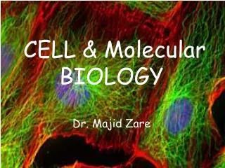
Cell Biology & Molecular Biology of The Cell. Lecturer Dr. Kamal E. M. Elkahlout , Assistant Professor of Biotechnnolgy Lecture 2 Protein Structure and Function Introduction to biotechnology registration for Biotech MSc. Protein Central Dogma.

E N D
Cell Biology & Molecular Biology of The Cell Lecturer Dr. Kamal E. M. Elkahlout, Assistant Professor of Biotechnnolgy Lecture 2 Protein Structure and Function Introduction to biotechnology registration for Biotech MSc
Protein Central Dogma • Proteins are large molecules that are formed as single, unbranchedchains of amino acid monomers • – But, proteins can be turned into branched structures by ubiquitinand other ubiquitin-like molecules • There are 20 different amino acids commonly found in proteins • • A protein’s amino acid sequence determines its three dimensional structure (conformation) • – Well, sort of…. • A protein’s 3-dimensional structure determines its chemical function(s) • – (along with a whole lot of different post-translational modifications that can alter parts of its structure and change its functions)
Fig. 2-14: The 20 common amino acids found in proteins.
The remaining amino acids have hydrophobic and “special” functional groups
Amino acids are linked by an amide linkage, called a peptide bond, to form polypeptide chains
Peptide bonds and the α carbon atoms form the linear backbone of proteins, which is a regular, repeating unit • The functional groups of amino acids form “side chains” that are connected to the backbone.
Polypeptide chains are flexible, butconformationally restricted
The shape of proteins is determinedthrough 4 levels of structure • Primary: the linear sequence of amino acids • Secondary: the localized organization of parts of a polypeptide chain (e.g., the α helix or β sheet) • Tertiary: the overall, three-dimensional arrangement of the polypeptide chain • Quaternary: the association of two or more polypeptides into a multi-subunit complex • The final, 3-dimensional, folded structure is generally one in which the free energy of the molecule is minimized
Three types of weak, non-covalent bonds also constrain the folding of proteins into their energy minimized 3-D structures
Hydrophobic interactions also playa role in determining protein shape • Residues with hydrophobic side chains tend to cluster in the interior of the protein molecule, avoiding contact with water • Polar side chains tend to be arranged on the outsides of proteins in contact with the aqueous medium
All of these bonds are about 30-300 times weaker than covalent bonds • So why are they important? • Many weak bonds applied together can produce a large force. • The stability of a protein is determined by the combined strength of many non-covalent bonds
Secondary Structure • The α-helix and the β-sheet are two regular folding patterns found in almost all proteins • What produces these structures and why are they so common? • They result from hydrogen bonding between multiple N-H and C=O groups in the backbone. • Side chains are not involved in these structures.
The α-helical backbone is a rigid cylinder with the amino acid side chains protruding from its surface
▲ FIGURE 3-2 Structure of a tripeptide. Peptide bonds (yellow) link the amide nitrogen atom (blue) of one amino acid (aa) with the carbonyl carbon atom (gray) of an adjacent one in the linear polymers known as peptides or polypeptides, depending on their length. Proteins are polypeptides that have folded into a defined three-dimensional structure (conformation). The side chains, or R groups (green), extending from the carbon atoms (black) of the amino acids composing a protein largely determine its properties. At physiological pH values, the terminal amino and carboxyl groups are ionized.
▲ FIGURE 3-3 The helix, a common secondary structure in proteins. The polypeptide backbone (red) is folded into a spiral that is held in place by hydrogen bonds between backbone oxygen and hydrogen atoms. The outer surface of the helix is covered by the side-chain R groups (green). Side chains protrude from the surface of the cylinder
α-helices can form very stable coiled-coil structures through hydrophobic interactions between non-polar side chains
β-sheets are found in the core ofmany proteins β-sheets are rigid, relatively flat and extended structures that are stabilized by hydrogen bonds between neighboring polypeptide strands
Secondary structure: the beta sheetWhere do the side chains go?
β-sheets can be in either a parallelor antiparallel orientation
Most extracellular proteins are stabilized by covalent –S-S- cross links Disulfide bond formation is catalyzed in the ER prior to export
Protein domains represent another important unit of organization
Figure 3-12. A protein formed from four domains. In the Src (tyrosine kinase involved in signaling between cells in multicellular animals) protein shown, two of the domains form a protein kinaseenzyme, while the SH2 and SH3 domains (Srchomolgydomain2 & 3) perform regulatory functions. (A) A ribbon model, with ATP substrate in red. (B) A spacing-filling model, with ATP substrate in red. Note that the site that binds ATP is positioned at the interface of the two domains that form the kinase.
Hierarchical Structure of Proteins • Domains are constructed from different combinations of α-helices and β-sheets at their core • Each combination is called a protein fold
Most large multi-domain proteins have evolved by recombination and joining of preexisting domains in new combinations (Domain Shuffling) • Many small molecule binding sites in proteins are created at the surfaces between new combinations of domains
Figure 3-18. Domain shuffling. An extensive shuffling of blocks of protein sequence (protein domains) has occurred during protein evolution. Those portions of a protein denoted by the same shape and color in this diagram are evolutionarily related. Serine proteases like chymotrypsin are formed from two domains (brown). In the three other proteases shown, which are highly regulated and more specialized, these two protease domains are connected to one or more domains homologous to domains found in epidermal growth factor (EGF; green), to a calcium-binding protein (yellow), or to a "kringle“ domain (blue) that contains three internal disulfide bridges.
Large proteins often contain more than one polypeptide chain • Binding between two protein surfaces generally involves sets of non-covalent bonds
Figure 3-21. Two identical protein subunits binding together to form a symmetric protein dimer. The Cro repressor protein from bacteriophage lambda binds to DNA to turn off viral genes. • Its two identical subunits bind head-to-head, held together by a combination of hydrophobic forces (blue) and a set of hydrogen bonds (yellow region).
Figure 3-22. A protein molecule containing multiple copies of a singleprotein subunit. The enzyme neuraminidase (glycoside hydrolasenz, neuraminin acid) exists as a ring of four identical polypeptide chains. The small diagram shows how the repeated use of the same binding interaction forms the structure.
Figure 3-23. A protein formed as a symmetric assembly of two different subunits. • Hemoglobin is an abundant protein in red blood cells that contains two copies of a globin and two copies of b globin. • Each of these four polypeptide chains contains a heme molecule (red), which is the site where oxygen (O2) is bound. • Thus, each molecule of hemoglobin in the blood carries four molecules of oxygen.
Some globular proteins can form long helical filaments • Globular proteins fold into a compact, ball-like shape with irregular surfaces • Example: Actin filaments form in a helical arrangement that can be the length of the cell
Proteins can be subunits for theassembly of large structures • enzyme complexes • ribosomes • Proteasomes (large proteases, degrade uneeded damage proteins) • filamentous structures (nuclear lamina) • viruses • membranes
Protein Function Some General Principles • All proteins bind to other molecules • Protein binding has a high degree of specificity for its ligands (binding partners) • Ligand specificity and affinity are determined by sets of weak non-covalent bonds and hydrophobic interactions.
Figure 3-38. The binding site of a protein. (A) The folding of the polypeptide chain typically creates a crevice or cavity on the protein surface. This crevice contains a set of amino acid side chains disposed in such a way that they can make noncovalent bonds only with certain ligands. (B) A close-up of an actual binding site showing the hydrogen bonds and ionic interactions formed between a protein and its ligand (in this example, cyclic AMP is the bound ligand).
Enzymes are highly specific catalysts • Enzymes speed reactions by selectively stabilizing unstable transition states (conformations) of their ligands • This lowers the activation energy of the reaction.
The catalytic activities of many enzymes are highly regulated through small molecule binding sites • Allosteric enzymes have two or more binding sites that interact with other molecules • – an active site that recognizes substrates • – a regulatory site that recognizes a regulatory molecule binding of a regulatory molecule at one site on the protein causes a conformational change in the polypeptide that can switch the active site conformation “On” or “Off”.
Figure 3-57. Positive regulation caused by conformational couplingbetween two distant binding sites. In this example, both glucose and moleculeX bind best to the closed conformation of a protein with two domains. Becauseboth glucose and molecule X drive the protein toward its closed conformation,each ligand helps the other to bind. Glucose and molecule X are therefore saidto bind cooperatively to the protein.