Download
1 / 54
551 likes | 796 Views
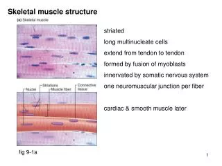
Skeletal Structure. Primary Functions of Bone. supports the soft tissues of the body so that the form of the body and an erect posture can be maintained protects delicate structures blood cell production - RBC, WBC, & platelets produced in red marrow

E N D
Primary Functions of Bone • supports the soft tissues of the body so that the form of the body and an erect posture can be maintained • protects delicate structures • blood cell production- RBC, WBC, & platelets produced in red marrow • storage for calcium & phosphorus; reserve lipid in yellow marrow • lever system with muscles-directs forces generated by muscles
Classified by Shape • long bones: longer than wide, shaft with 2 ends (femur, humerus) • short bones: length equals width (carpals, tarsals) • flat bones: thin and flat (cranium, sternum, ribs, scapula) • irregular bones: complex shapes (vertebrae, middle ear bones) • sesamoid bones: small bones formed in tendons (patella)
Structure • diaphysis: shaft; epiphyseal line separates it from the epiphysis • epiphysis: end; contains red bone marrow • articular cartilage: hyaline cartilage on joint surfaces to resist friction
Structure (cont) • periosteum: dense, white, fibrous covering around the remaining surface of the bone • outer layer: composed of connective tissue containing blood vessels • inner osteogenic layer: contains elastic fibers, blood vessels, and osteoblasts (cells responsible for forming new bone during growth and repair) • medullary cavity: space within the diaphysis that contains the fatty yellow marrow in adult bones
Structure (cont) • endosteum: layer of osteoblasts that lines the medullary cavity and contains scattered osteoclasts (cells that may assume a role in the removal of bone) • nutrient foramen: opening allowing vessels to enter the medullary cavity supplying the shaft of the bone (arteries to the epiphysis generally arise from the joint capsule)
Bone Density • Compact bone: dense bone of the diaphysis consisting of repeating patterns of solid bone tissue organized into concentric layers (long bones) • Spongy or cancellous bone: latticework (large spaces) arrangement packed with red marrow; resists stresses of weight & postural changes as well as muscular development (short, flat, irregular, and epiphysis of long bones)
Classification of Joints • Fibrous (synarthroses): lacks a joint cavity and the articulating bones are held very closely together by fibrous connective tissue; they permit little or no movement • sutures • syndesmoses • gomphoses
Sutures • found between the bones of the skull and are united by a thin layer of dense fibrous connective tissue
Syndesmoses • fibrous connective tissue forms an interosseous membrane or ligament (distal articulation of the tibia and fibula, shafts of radius and ulna)
Gomphoses • cone-shaped peg fits into a socket (teeth)
Classification of Joints • Cartilaginous (amphiarthroses): lacks a joint cavity and the articulating bones are tightly connected by cartilage • synchrondrosis • symphyses
Synchrondrosis • connecting material is hyaline cartilage (epiphyseal plate)
Symphyses • connecting material is a broad, flat disc of fibrocartilage (pubic symphysis; bodies of vertebrae)
Classification of Joints • Synovial (diarthroses): joint cavity (space between the articulating bones) is present; freely movable. • Gliding • Hinge • Pivot • Ellipsoidal • Saddle • Ball and socket
Gliding • side-to-side and back-and-forth movements (biaxial); articulating surfaces are usually flat (intercarpal, intertarsal, sternum and clavicle)
Hinge • motions are flexion/extension (monoaxial); convex surface of one bone fits into the concave surface of another (elbow, knee)
Pivot • rotational movement (monoaxial); rounded, pointed, or concave surface fits into a ring formed partly by bone and partly by a ligament (atlas and axis)
Ellipsoidal • side-to-side and back-and-forth movements (biaxial); oval shaped condyle fits into an elliptical cavity (wrist)
Saddle • side-to-side and back-and-forth movements (biaxial); articular surfaces concave in one direction and convex in opposite direction (CMC of thumb)
Ball and socket • movement in 3 planes (triaxial); ball like surface fits into a cuplike depression (shoulder and hip)
Components of Synovial Joints • articular cartilage: covers surfaces of articulating bones but does not bind them together • articular capsule: surrounds the articular surfaces and encloses the joint cavity • outer layer (fibrous capsule): attached to the periosteum of articulating bones at a variable distance from the edge of the articulating cartilage • inner layer (synovial membrane): secretes synovial fluid which lubricates the joint and provides nourishment for the articular cartilage
Components of Synovial Joints • joint (synovial) cavity: enclosed space that surrounds the 2 articulating surfaces; contains the slippery lubricating fluid called synovial fluid • ligaments: thickened collagenous bands connecting bone to bone • extracapsular ligaments are outside of the articular capsule (MCL, LCL) • intracapsular ligaments directly attach the 2 articulating surfaces (ACL, PCL)
Components of Synovial Joints • articular discs (menisci): pads of fibrocartilage that lie between the articular surfaces of the bones; help maintain the stability of a joint and direct the flow of synovial fluid to areas of greatest friction; not all synovial joints have them
Components of Synovial Joints • bursae: saclike structures that contain synovial fluid to help reduce friction between: • skin and bone • tendons and bones • muscles and bones • ligaments and bones
Body planes • midsagittal: divides body down the middle • frontal (coronal): divides body to front and back • transverse: divides body into top and bottom half
transverse midsaggital coronal
Anatomical directions • superior: nearer the head • inferior: nearer the feet • lateral: away from the midline • medial: towards the midline
Anatomical directions • anterior: toward the front • posterior: toward the rear or back • proximal: nearer to the center • distal: farther from the center
Joint Movements • Flexion: decrease in the angle between the anterior (may be posterior--knee) surfaces of the articulating bones • Extension: increase in the angle between the anterior (may be posterior--knee) surfaces of the articulating bones • Hyperextension: continuation of extension beyond the anatomical position
Joint Movements • Dorsiflexion: flexion of the foot at the ankle joint • Plantarflexion: extension of the foot at the ankle joint
Joint Movements • Inversion: movement of the sole of the foot inward at the ankle joint • Eversion: movement of the sole of the foot outward at the ankle joint
Joint Movements • Abduction: movement of a bone away from the midline of the body • Adduction: movement of a bone toward the midline of the body
Joint Movements • Internal (medial) rotation: rotary motion in the transverse plane toward the midline • External (lateral) rotation: rotary motion in the transverse plane away from the midline • Circumduction: movement in which the distal end of a bone moves in a circle while the proximal end remains stable
Joint Movements • Elevation: movement in which a part of the body moves upward • Depression: movement in which a part of the body moves downward
Joint Movements • Protraction: movement forward on a plane parallel to the ground • Retraction: movement backward on a plane parallel to the ground