Download
1 / 69
700 likes | 988 Views
Bone Structure and Skeletal System. Chapters 6, 7, and 8. Function of the Skeletal System. The skeletal system has five functions. Support Storage of minerals and lipids Blood cell production Protection Leverage. Support.

E N D
Bone Structure and Skeletal System Chapters 6, 7, and 8
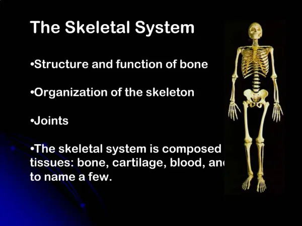
Function of the Skeletal System • The skeletal system has five functions. • Support • Storage of minerals and lipids • Blood cell production • Protection • Leverage
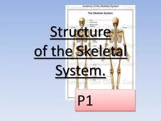
Support • The skeletal system provides structural support for the entire body. • Individual bones or groups of bones provide a framework for the attachment of soft tissues and organs.
Storage of minerals and lipids • Calcium is the most abundant mineral in the human body. • Calcium salts of bone are a valuable mineral reserve that maintains normal concentrations of calcium and phosphate ions in the body fluids. • Also acts as a mineral reserve, the bones of the skeleton store energy reserves as lipids in areas filled with yellow marrow
Blood Cell Production • Red blood cell, white blood cells, and other blood components are produced in red marrow which fills the internal cavities of many bones.
Protection • Many soft tissues and organs are surrounded by skeletal elements. • The ribs protect the heart and lungs, skull encloses the brain, the vertebrae shields the spinal cord, and the pelvis cradles delicate digestive and reproductive organs.
Leverage • Many bones function as levers that can change the magnitude and direction of the forces generated by the skeletal muscles. • The movements produced range from dainty motion of the fingertips to changes in the position of the entire body.
Cellular structures of Bones • The process of bone formation is osteogenesis. This is done with: • Osteoblast- immature bone cells; mononucleate bone-forming cells, that also manufacture hormones, and eventually become entrapped in the bone matrix to become osteocytes. • Osteocytes-the mature bone cell, they occupy are known as lacunae; functions include: formation of bone, matrix maintenance and calcium homeostasis. Also its been shown to act as mechano-sensory receptors — regulating the bone's response to stress and mechanical load.
Cellular structures of Bones • Osteolysis is the breaking down of bones • Osteoclast- are the cells responsible for bone resorption, they break down bone; the cells are large, and multinucleated they are located on bone surfaces equipped with phagocytic-like mechanisms that activate enzymes to breakdown bone
Bone Tissue • a specialized form of connective tissue and it is the main element of the skeletal tissues • It is composed of cells and an extracellular matrix in which fibers are embedded. • Bone tissue is unlike other connective tissues in that the extracellular matrix becomes calcified.
Bone Shapes • Every adult skeleton contains 206 major bones, which can be divided into six broad categories according to their individual shapes. • Long bones • Flat bones • Sutural bones • Irregular bones • Short bones • Sesamoid bones
Long Bones • Long bones are relatively long and slender. • Long bones are located in the arm, forearm, thigh, leg, palms, soles, fingers and toes. • The femur, the long bone of the thigh, is the largest and heaviest bone in the body.
Flat Bones • Flat bones have thin, roughly parallel surfaces. Flat bones form the roof of the skull, the sternum, the ribs, and scapula. • They provide protection for underlying soft tissue, and offer extensive surface area for the attachment of skeletal muscles.
Sutural Bones • Sutural bones or Wormianbones, are small, flat, irregularly shaped bones between the flat bones of the skull. • There are individual variations in the number, shape and position of the sutural bones. Their borders are like pieces of a jigsaw puzzle, and they range in size from a grain of sand to a quarter.
Irregular Bones • Irregular bones have complex shapes with short, flat notched, or ridge surfaces. • The spinal vertebrae, the bones of the pelvis, and several skull bones are irregular bones.
Short Bones • Short bones are small and boxy. • Examples of short bones include the carpal bones (wrists) and tarsal bones (tarsal)
Sesamoid Bones • Sesamoid bones are generally small, flat, and shaped somewhat like a sesame seed. • They develop inside tendons and are most commonly located near joints at the knees, the hands, and feet. • Sesamoid bones may form in at least 26 locations.
Bone MarkingsSurface Features • Surface features can yield an abundant amount of anatomical information. • Anthropologists, criminologist, and pathologist can often determine the size, age, sex and general appearance of an individual on the basis of incomplete skeletal remains.
Surface Features • Elevations and projections(general): • Process: any projection or bump • Ramus: an extension of a bone making an angle with the rest of the structure.
Surface Features • Processes formed where tendons and ligaments attach. • Trochanter: large, rough projection. • Tuberosity: a smaller, rough projection. • Tubercle: a small, rough projection • Crest: a prominent ridge • Line: a low ridge • Spine: a pointed process
Surface Features • Processes formed for articulation with adjacent bones. • Head: expanded articular end of an epiphysis, separated from the shaft by a neck. • Neck: narrow connection between the epiphysis and diaphysis
Surface Features • Condyle: a smooth, rounded articular process • Trochlea: a smooth, grooved articular process shaped like a pulley. • Facet: a small, flat articular surface
Surface Features • Depressions • Fossa: a shallow depression. • Sulcus: a narrow groove
Surface Features • Openings • Foreman: a rounded passageway for blood vessels or nerves. • Canal: a passageway through the substance of a bone. • Fissure: an elongated cleft. • Sinus or antrum: a chamber within a bone, normally filled with air
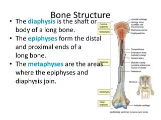
Bone Structure • Diaphysis: shaft of a long bone • Epiphysis: the head if a long bone • Metaphysis: the region of the long bone between the epiphysis and diaphysis, corresponding to the location of the epiphyseal cartilage of the developing . • Marrow cavity: or medullary cavity the space within in the bone that contains the marrow. • Cortex: Spongy bone that consists of an open network of struts and plates with a thin covering of compact bone.
Bone HistologyImportant Vocabulary The matrix of bone is very dense and contains deposits of calcium salts. Osteocytes: A bone cell responsible for the maintenance and turnover of the mineral content of the surrounding bone. They account for most of the cell population Lacuna: small pit or cavity Canaliculi: Microscopic passage way between cells: in bone canaliculi permit the diffusion of nutrients and waste to and from the osteocytes. Periosteum : the layer surrounding the bone, consisting of an outer fibrous region and an inner cellular region. Lamellae: concentric layers; the concentric layers of bone within an osteon.
Important Vocabulary Osteoblast: a cell that produces the fibers and matrix of the bone. In a process called osteogenesis. Osteoid: organic matrix of the bone before calcium salt are added. Osteoclast: a cell that dissolves the fiber and matrix of the bone. Osteogenic layer: the inner, cellular, layer of periosteum that participates in bone growth and repair. Osteolysis: the breakdown of the mineral matrix of the bone. Osteon: the basic histological unit of compact bone, consisting of osteocytes organized around a central canal and separated by concentric lamellae.
Structure of Compact Bone The basic functional unit of mature compact bone is the osteon, or Haversian System. In an osteon, the osteocytes are arranged in concentric layers around a central canal, or Haversian canal.This canal contains one or more blood vessels that carry blood to and from the osteon. Central canals generally run parallel to the surface of the bone.
Other passageways, known as perforating canals or canal of Volkmann, extend roughly perpendicular to the surface. Blood vessels in these canals supply to osteons deeper in the bone to tissue of the marrow cavity.
The Structure of Spongy Bone In spongy bone the lamellae are not arranged in osteons. The matrix in spongy bone forms struts and plates called trabeculae. The thin trabeculae branch, creating an open network. There are blood vessels in the matrix of the spongy bone. Nutrients reach the osteocytes by diffusion along the canaliculi that open onto the surface of trabeculae. Red marrow is found between the trabeculae of spongy bone, and blood vessels within the tissue delivers nutrients to the trabeculae and remove waste generated by the osteocytes
Spongy bone is located where bones are not heavily stress or where stresses arrive from many directions. Spongy bone is much lighter than compact bone. Finally, the framework of trabeculae supports and protects the cells of the bone marrow. Red marrow is responsible for blood cell formation and yellow bone marrow- adipose tissue is important for energy reserves.
Periosteum Periosteum: The layer that surrounds a bone, consisting of a an outer fibrous region and inner cellular region. 1.periosteum isolates the bone from surrounding tissue. 2. provides a route for circulatory and nervous supply. 3. actively participates in bone growth and repair. Near the joints the periosteum becomes continuous with the connective tissues that lock the bones together. The fibers of the periosteum are interwoven with those of tendons attached to the bone.
Endosteum Endosteum: an incomplete layer that lines the marrow cavity. This is the layer which is active during bone growth, repair, and remodeling, covers
Bone Growth Formation The boney Skelton begins to form about 6 weeks after fertilization, when the embryo is approximately 12 mm long. At this stage the existing skeleton elements are nothing more than cartilaginous.
The process of replacing tissues with bone is called ossification. This term refers specially to the formation of bone. The process of calcification - the deposition of calcium salts – occurs during ossification.
Most simply the process of bone formation or ossification involves two major phases. 1st step: hyalinecartilage model is completely covered with bone matrix (bone collar) by bone forming cells called osteoblasts.
By birth or shortly after the hyaline cartilage models have been converted to bone except for two regions: articular cartilages(that cover the bone ends) and the epiphysealplates. Thearticular cartilages persist for life, reducing friction at the joint surfaces. The epiphyseal plates provide for longitudinal growth of the long bones during childhood. During development, most bones originate as hyaline cartilage that are miniature models of the corresponding bones of the adult skeleton.
Endochondral Ossification Cartilage is replaced by bone Examples: most bones of the body Involves a 6-step process: 1. A cartilage model forms 2. Growth occurs by interstitial & appositional mechanisms 3. Primary ossification centers develop 4. A medullary cavity develops 5 Secondary ossification centers develop at epiphyses 6. Hyaline cartilage is replaced by articular cartilage at the ends, and between the diaphysis and the epiphyses by the epiphyseal (bony) plate.
Intramembranous Ossification Simpler process than endochondral ossification. Examples: flat bones of the skull Involves a 4-step process: 1. An ossification center develops 2. Calcification occurs due to mineral deposition 3. Trabeculae are formed in the interior 4. Mesenchyme is replaced with periosteum and a thin layer of compact bone
The Blood and Nerve Supplies Osseous tissue is highly vascular, and the bones of the skeleton have an extensive blood supply. In a typical bone such as the humerus, three major sets of blood vessels develop. 1. Nutrient Artery and Vein: the blood vessels that supply the diaphysis form by invading the cartilage model as endochondral ossification begins. The vessels enter the bone through one or more passageways called nutrient foramina in the diaphysis
Metaphseal Vessels: supply blood to the inner (diaphyseal) surface of each epiphyseal cartilage is being replaced by bone. Periosteal Vessels: Blood vessels from the periosteum provide blood to the superficial osteons of the shaft. During endochondral formation, branches of periosteal vessels enter the epiphyses, providing blood to the secondary ossification centers.
Dynamic Nature of Bones The organic and mineral components of the bone matrix are continuously being recycled and renewal through the process of remolding. Bone remolding goes on throughout life, as part of normal bone maintenance. Remolding can replace the matrix but leave but leave the bone as a whole unchanged, or it may shape, internal architecture, or mineral content of the bone.
Bone is continually remodeled, recycled, and replaced. The rate of turnover varies from bone to bone and from moment to moment. When deposition exceeds removal, bone gets stronger; when removal exceeds deposition, bones get weaker.
Effects of Exercise on the bone Turnover and recycling of minerals give each bone the ability to adapt to new stresses. The sensitivity of osteoblasts to electrical events theorized as the mechanism that controls the internal organization and structure of bone.
When bone is stressed, the mineral crystals generate minute electrical fields. Osteblasts are apparently attracted to these electrical fields and, once in the area, begin to produce bone. This finding has led to the successful use of small electrical fields in stimulating the repair of severe fractures. When you don’t use you lose. The stresses applied to the bones during physical activity are essential to maintaining bone strength and bone mass.