Download
1 / 47
470 likes | 475 Views
Learn about the structures, functions, and divisions of the human nervous system. Understand the sensory and motor pathways and the functional division of the peripheral nervous system. Explore the autonomic nervous system and its role in maintaining a stable internal environment.

E N D
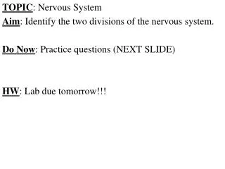
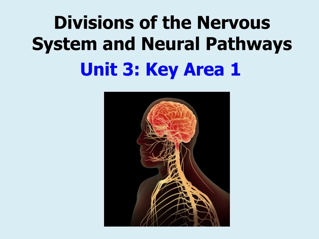
Divisions of the Nervous System and Neural Pathways Unit 3: Key Area 1
Produce a spider diagram on the back of your hand out to summarise everything you already know about the….. • Once you have finished share with others in your group and add to your diagram. nervous system
Learning Intentions • Describe the structures and functions of the human nervous system.
The Nervous System • The nervous system analyses sensory information from the body and the external environment, stores some aspects and makes decisions regarding appropriate responses and behaviours. • It makes motor responses by initiating muscular contractions and glandular secretions.
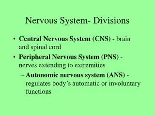
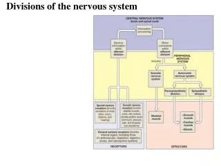
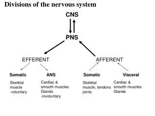
Structural Division of the nervous system • The nervous system consists of the brain, spinal cord and peripheral nerves. • It can be divided into the central nervous system (CNS) and the peripheral nervous system (PNS). Central nervous system The CNS is made up of the brain and spinal cord.
Structural Division of the nervous system Peripheral nervous system • The PNS consists of all parts of the nervous system except for the CNS. • It includes all the cranial and spinal nerves and their branches which link to the receptors and effectors.
Structural Division of the nervous system • In summary: brain Central nervous system (CNS) Spinal cord Nervous system Peripheral nervous system (PNS) Cranial nerves Spinal nerves
Sensory&MotorPathways • Many of the peripheral nerves contain a sensory pathway consisting of sensory neurones and/or a motor pathway consisting of motor neurones. Motor pathways carry impulses from the CNS to effectors Sensory pathways carry impulses from receptors in the sense organs to the CNS CNS (brain & spinal cord) Nerve impulses carried by sensory neurones Nerve impulses carried by motor neurones External e.g. skin, eye retina Internal e.g. CO2 receptors, thermoreceptors receptors e.g. muscles, glands effectors e.g. limb movement; hormone production e.g. sounds, sights, temperature of skin/blood etc stimuli response
FunctionalDivision of the nervous system • The nervous system can also be divided according to functions as well as structurally.
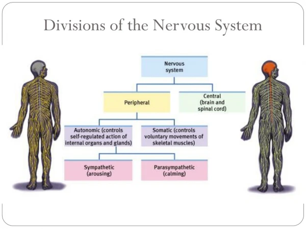
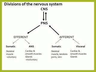
FunctionalDivision of the nervous system Peripheral nervous system (PNS) The overall function of the PNS can be split into two functional parts: • Those that are controlled in a voluntary way by the somatic nervous system (SNS) acting through skeletal muscles. • Those that are controlled in an involuntary way by the autonomic nervous system (ANS) acting through smooth muscles, cardiac muscle and glands. Somatic nervous system Autonomic nervous system
FunctionalDivision of the nervous system Somatic nervous system (SNS) • In brief: Peripheral nervous system (PNS) Autonomic nervous system (ANS) sympathetic parasympathetic
Somatic nervous system (SNS) • The somatic nervous system (which includes spinal nerves) controls the body’s skeletal muscles. • This involves sensory and motor pathways as outlined on the flow diagram on an earlier slide (see opposite). • The somatic nervous system is responsible for bringing about certain involuntary reflex actions (e.g. limb withdrawal) but most of the control that it exerts is over voluntarymovements of the skeletal muscles.
The Autonomic nervous system (ANS) • The autonomic nervous system normally works automatically without the person’s conscious control. • It controls functions that bring about a steady internal state within the body. These functions include regulating: • Heart rate • Breathing rate • Intestinal secretions • The ANS is composed of sympatheticand parasympatheticsystems that work to regulate these processes.
The Autonomic nervous system (ANS) Examine the diagram on the slide which follows.... Think about.... Where does the autonomic nervous system originate? Why is it describes as being antagonistic? What do you notice about the adrenal gland?
The Autonomic nervous system (ANS) • Harmonious balance... • The autonomic nervous system is concerned with maintaining a stable internal environment by playing its part in the process of homeostasis. • The autonomic nervous system originates in the medulla of the brain. • The sympathetic and parasympathetic systems are described as being antagonistic because they have oppositeeffects on the same body structures. • There is no parasympathetic nerve connected to the sweat glands or the adrenal glands. • To return the sweat and adrenaline production back to normal levels the number of nerve impulses to the sympathetic nerves are reduced.
Antagonistic in Action The sympathetic system prepares the body for fight or flight. The parasympathetic system prepares the body for rest and digest. Overall, the two divisions work together to ensure that the body responds appropriately to different situations.
Effects of the sympathetic and parasympathetic systems rate and force of contraction decreases Heart Smooth muscle in bronchiole wall relaxes rate of peristalsis and intestinal secretions increases Digestive Tract
Summary of Nervous System Structure & Function Key… structure function Here is one way of summarising the nervous system…
HUMAN NERVOUS SYSTEM- Classified according to function….) Responsible for certain involuntary actions e.g. reflex actions involving movement such as limb withdrawal. Autonomic • ___________ nervous system • (ANS) • ______________ • Regulates internal structures and organs e.g. heart, lungs etc. • Somatic • nervous system (SNS) • Mostly voluntary • Controls: involuntary Skeletal muscles - Classified according to structure Sympathetic System __________________ __________________ e.g. increases heart rate Parasympathetic system Calms the body down returning it to its normal state e.g. _______________ _______________ Prepares body for action CENTRAL nervous system PERIPHERAL nervous system Reduces heart rate Cranial & peripheral nerves Brain and Spinal cord
Past paper question • The diagram below shows how the nervous system is organised: • Complete the diagram by entering the names of parts A to D. (2) • (b) The parts of the autonomic nervous system are described as • antagonistic. • (i) What is meant by the term antagonistic? • __________________________________________ (1) • (ii) Explain how this antagonistic action controls the activity of • the digestive system. • _______________________________________________________________________________________________________________________________________________________________________________ (2) Nervous system A. somatic Autonomic system B. CNS Peripheral system sympathetic Parasympathetic D. Spinal cord C. Brain They have opposite effects (on the same organ) e.g. Peristalsis is stimulated by the parasympathetic system OR - intestinal secretions are stimulated by the parasympathetic system OR - Blood flow is stimulated by the parasympathetic system (1mk) Parasympathetic stimulates the digestive system, whereas the sympathetic system inhibits the digestive system (1mk) 2000
Extended Answer Question • Describe the structure and function of the autonomic nervous system. (7)
Marking Scheme ANS works automatically / without conscious control Impulses originate in the medulla (region of the brain) It is made up of the sympathetic andparasympathetic systems These two systems are antagonisticin action The sympathetic system prepares the body for fight or flight The parasympathetic system prepares the body for rest and digest Correct description of the effect of the ANS in controlling heart rate Correct description of the effect of the ANS in controlling breathing rate Correct description of the effect of the ANS in controlling peristalsis Correct description of the effect of the ANS in controlling intestinal secretions
The Cerebral Cortex Key Area 2
Learning Intentions • Describe the structures and functions of the human brain.
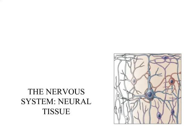
The Human Brain • The brain is a complex central processing centre of the nervous system. • It receives incoming information from sense organs and responds by initiating appropriate actions. • It is made up of billions of neurons.
The Cerebral Cortex • The cerebrum consists of a thick outer surface called the cerebral cortex (3mm deep) which forms the largest and most complex part of the brain.
Cerebral Cortex: Function • The cerebral cortex is involved in consciousactions such as: • Receiving sensory information • Co-ordinating Voluntary movement • Making decisions in light of experience
Convoluted Surface • The cerebral cortex is highly convoluted (folded in on itself) to increase its surface area which…. • allows a large number of cell bodies (of neurons) to be located on the surface. (i.e. to allow space for more nerve cells) • increases the number of interconnections between neurons; (i.e. to allow space for more nerve cell connections)
Past Paper Qu Explain how the maximum number of interconnections between neurones is achieved within the cerebrum. (2mk) (The cerebrum) has a convoluted/folded surface/large surface (1mk) This allows for an increased number of cell bodies/ cells/neurones (1mk)
The Cerebral Cortex • The cerebrum is split by a deep cleft into two halves called cerebral hemispheres. • The lefthemisphere processes information from the right visual field and controls the rightside of the body (the reverse is true for the right hemisphere).
Corpus callosum The two cerebral hemispheres are connected by a large bundle of nerve fibres called the corpus callosum. -This structure transfers information in the form of impulses between the two sides of the brain. Corpus callosum
Location of the Corpus callosum 1 2 Cross section of cerebrum Horizontal section of cerebrum 3 4 5
Functional Areas of the Cerebral Cortex • The cerebral cortex can be divided into 3 types of functional area: • - Sensory • - Association • - Motor • Each area is discrete– this means it performs its own particular function, distinct from the other areas.
Functional Areas of the Cerebral Cortex Sensory area – receives information from body receptors e.g. touch receptors in the skin; thermoreceptorsin the hypothalamus. Association area – Analyses and interprets these impulses from the body's receptors and makes sense of them. Makes decisions if necessary. There are association areas involved in language processingandpersonality, imagination and intelligence. Motor area – receives information from the association areas and carryout ‘orders’ by sending motor impulses to the effectors e.g. muscles; glands.
Related Topic Motor Area (aka ‘motor strip’) The motor area is one of the largest regions of each cerebral hemisphere. Each motor area consists of motor neurones which send out impulses to bring about voluntarymovement of skeletal muscles. However, the size of the part of the motor area devoted to any one part of the body operated by skeletal muscles is not in proportion to the actual size of the body part. Instead the amount of motor area is found to be in proportion to the relative number of motor endings present in that body part (i.e. mobility/fine motor control).
Related Topic Motor homunculus The diagram opposite shows…….. an imaginary human figure whose body parts have been drawn in proportion to their mobility and fine motor controlas opposed to their actual size. Note the large amount of the motor area devoted to mobility of the lips and hands.
Related Topic Sensory Area (aka sensory ‘strip’) •Receives impulses from the sense organs of the body to allow exact location of a sensation e.g. a pin prick. •large areas are devoted to parts of the body which are more sensitive than others such as the hands, lips and tongue. Sensory homunculus model… Parts of the body are sized according to how much space the brain gives to processing sensory information about that part of the body.
Learning Intentions • Revise some of the key points about the brain by producing a visualiser and trying practice question/s. • To investigating some interesting topics about the brain and share our findings with other group members.
Make a visualiser to summarise key points about the brain… 3 functional areas: Sensory Association Motor How does yours compare to this example…? BRAIN central core Limbic system Cerebral cortex highly convoluted hypothalamus cerebellum medulla • long-term memories • emotional states biological motivation • Receiving sensory information • Co-ordinates voluntary movement • Making decisions in light of experience • Personality, imagination, intelligence, thought etc. Controls balance, posture and co-ordinates movement Regulates e.g. breathing, heart rate, peristalsis Outer surface of cerebrum Right side receives info from left visual field and controls left side of body (opposite for left side) Cerebrum made up of two cerebral hemispheres Corpus callosum: bundle of nerve fibres connect the hemispheres and transfers information between two hemispheres
Past Paper question The diagram shows the main parts of the human brain as seen in a vertical section. thought, reasoning, personality, imagination, intelligence, recalls memories, motor control, shapes behaviour, receives sensory information etc. (a) Complete the table by adding the correct letters, names and functions. Cerebral cortex P S Pituitary gland R medulla Regulates sleep Hormone production/any named hormone (b) Describe a feature of part P which improves its function. its large surface area/its folded/convoluted nature (c) What is meant by the term “localisation of function”? Specific areas of the brain are involved in specific tasks (d) Explain the difference between somatic and autonomic actions. Somatic actions are voluntary and brought about by the skeletal muscles - 1mk Autonomic actions are involuntary and brought about by smooth muscle/cardiac muscle/glands - 1mk 2
Brain Studies • Expert Jigsaw: Research topics • Evidence from Brain Injuries • Electroencephalograms (EEGs) • Split Brain Studies • Imaging the brain (brain scans) • Research your topic using pages 218-222 of your textbook and the information pack your teacher gives you. • Be ready to discuss/report key points with your group members.
Video clip: Brain Scans Vegetative state patients can respond to questions http://news.bbc.co.uk/1/hi/health/8497148.stm(2:38) Ed Scotland clip http://www.educationscotland.gov.uk/highersciences/humanbiology/unitthree/nervoussystem/index.asp (3:29)
Video clips: Split Brain Patients YouTubevideos of split brain patients... ** Severed corpus callosum (10.11) ** Split brain behavioural experiments (4.35) ** Curious: Split brain (14.23) (rare condition were people are born without their corpus callosum).
Try this…. The composite picture on the left was shown to several split-brain patients. A little while later they were asked to study the pictures on right. State, with reasons, which picture the patient will choose when asked to (i) say what they had seen; (ii) point with their left hand to what they had seen. • When asked to say what they had seen: They choose B – the man’s face. This is because the right side of the image (man’s face) becomes an image on the left side of the brain which is where speech is located. (ii) When asked to point with their left hand: They choose C – the woman’s face. This is because the left side of the image (woman’s face) becomes an image on the right side of the brain and the right side of the brain controls the left hand.
Past Paper question corpus callosum Try 2013 Qu 11 The patient sees “racket” and can say the word because the left side of the brain controls language production (1mk) Patient sees “tennis” but cannot say the word because the link to the left side of the brain/language production centre is lost (1mk)