Download
1 / 57
580 likes | 616 Views
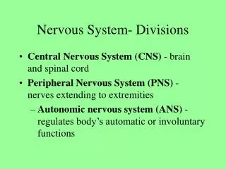
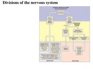
Divisions of the nervous system. CNS. PNS. EFFERENT. AFFERENT. Somatic. ANS. Somatic. Visceral. Cardiac & smooth muscles Glands -involuntary. Cardiac & smooth muscles Glands. Skeletal muscle -voluntary. Skeletal muscle, tendons joints, skin. PNS Terminology.

E N D
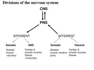
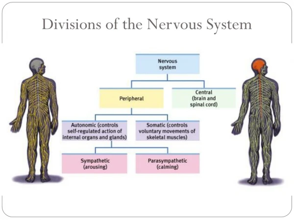
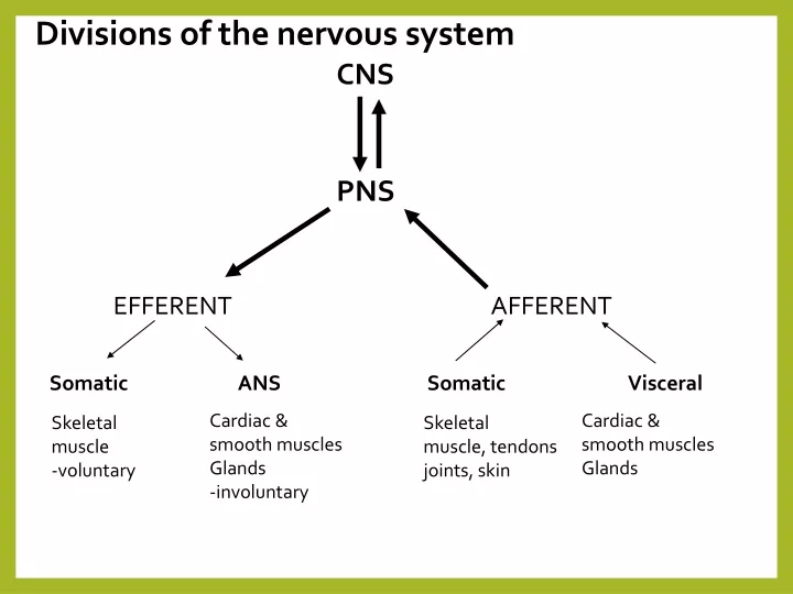
Divisions of the nervous system CNS PNS EFFERENT AFFERENT Somatic ANS Somatic Visceral Cardiac & smooth muscles Glands -involuntary Cardiac & smooth muscles Glands Skeletal muscle -voluntary Skeletal muscle, tendons joints, skin
PNS Terminology • Ganglia = neuron cell bodies • Peripheral nerves = neuronal axons • PNS neuroglia • Satellite cells • Enclose neuron cell bodies in ganglia • Schwann cells • Cover peripheral axons
Ganglion • plural = ganglia • collection of neuronal cell bodies • neuron leading into a ganglion = pre-ganglionic neuron • cell bodies are located in the CNS (brain or spinal cord) • axons synapse with the cell bodies of the ganglion • the cell bodies and axons leading from the ganglion = post-ganglionic neuron
Efferent Division of the PNS • the somatic nervous system and part of the autonomic nervous system • the somatic = control of skeletal muscle • the ANS = involuntary control over cardiac and smooth muscle + gland secretion
I - Olfactory II - Optic III - Oculomotor IV-Trochlear V - Trigeminal VI - Abducens VII - Facial VIII - Acoustic IX - Glossopharyngeal X - Vagus XI - Accessory XII - Hypoglossal -cranial nerves – 12 pairs -considered part of the peripheral nervous system (PNS) -olfactory & optic contain only sensory axons = sensory nerves -remaining are motor or mixed nerves (both motor and sensory axons)
Dorsal Root of SN Ventral Root of SN SPINAL NERVE Dorsal Ramus Ventral Ramus Rami Communicantes Sensory – IN Motor – OUT SKIN of BACK BACK MUSCLES Sensory – IN Motor – OUT TRUNK LIMBs (including skin) Signals to and from the ANS VISCERA – cardiac and Smooth muscle
Spinal Nerve • after passing through intervertebral foramina the spinal nerve branches into 3 rami (singular ramus) • 1. Dorsal ramus • Sensory/motor innervation to skin and muscles of back • 2. Ventral ramus • Sensory/motor innervation to ventral and lateral body surface, body wall structures, muscles of the upper and lower limbs
3. rami communicantes = a connection between a spinal nerve and the sympathetic trunk of the ANS • two types: • gray ramus communicantes – unmyelinated post-ganglionic axons • white ramus communicantes – myelinated preganglionic axons
Somatic Nervous System • somatic/motor axons emerge from the ventral gray horn and travel into the spinal nerve • they then travel through either the: • dorsal ramus to end up at the muscles of the back • OR the ventral ramus to end up at the muscles of the limbs and body wall (chest/abs/pelvis)
Somatic Nervous System • considered the voluntary aspect of the PNS • but the muscles of posture and balance are controlled involuntarily by the lower brain centers (brain stem, cerebellum) • cell bodies located in the ventral gray horn of the spinal cord or nuclei of the brain stem • the axons extend continuously to its skeletal muscle target • synaptic terminals release acetylcholine – contraction of skeletal muscle • can only stimulate its target
Somatic Nervous System • somatic motor neurons originate in the ventral gray horn or the brain stem • receive incoming information from many presynaptic neurons • both excitatory and inhibitory on the somatic motor neurons
Somatic Nervous System • somatic motor neurons also synapse with: • 1. reflex interneurons originating in the spinal cord • 2. upper level neurons from motor areas of the brain – form the descending white matter tracts • these neurons synapse with the somatic motor neurons and regulate their activity • activation – impulse sent to muscles • inhibition – no impulse, no contraction
Somatic Nervous System • no matter what motor pathway you learn – they eventually affect the somatic motor neuron that originates in the ventral gray horn or the brain stem • therefore the somatic motor neuron is considered the final common pathway • considered the only way any other part of the nervous system can influence muscle activity
Somatic Motor pathways • all excitatory and inhibitory signals that control skeletal muscle movement converge on the somatic motor neurons • these somatic motor neurons originate in one of two places: • 1. Brain Stem nuclei • 2. Ventral Gray Horn of Spinal Cord • these somatic motor neurons extend from the brain stem and SC to innervate the skeletal muscles • they are also called lower motor neurons (LMNs) • their axons travel via cranial and spinal nerves to skeletal muscle • only LMNs provide output from the CNS to skeletal muscle fibers • damage to the LMNs produces flaccid paralysis on the same side as the damage – loss of reflex action, motor tone and voluntary contraction
Somatic Motor pathways • neurons in four distinct circuits control movement by providing input to these LMNs • 1. local circuit neurons • 2. upper motor neurons (UMNs) • 3. basal ganglial neurons • 4. cerebellar neurons
UMNs: Upper Motor Neurons • provide input to the local circuit and LMNs • essential for planning, initiating and directing sequences of voluntary movements • extend from the brain to the LMNs via two types of somatic motor pathways: • 1. Direct Pathways • 2. Indirect Pathways
UMNs: Upper Motor Neurons • 1. direct motor pathways: nerve impulses for voluntary movement • lateral corticospinal, anterior corticospinal and corticobulbar (brain stem) • UMNs originate in the motor cortex and travel down the spinal cord as the corticospinal tracts to synapse with the LMN • OR – UMNs exit the brain stem as corticobulbar tracts • the LMN emerges as spinal nerves or through the brain stem and out as cranial nerves
Direct Motor Pathway: The Corticospinal tracts • 1. lateral corticospinal • 2. anterior corticospinal • major motor tract for voluntary skeletal muscle movement • especially fine motor skills • UMNs originate from motor cortex and travel through brain stem • as they pass through the medulla they form the pyramids • lateral corticospinal – one tract decussates; the other continues • connection between UMN and LMN may involve a local circuit interneuron • e.g. lateral corticospinal to LMN
UMNs: Upper Motor Neurons • 2. indirect motor pathways: or extrapyramidal pathways • nerve impulses follow complicated circuits that involve the cortex, basal ganglia, thalamus and brain stem • descending axons/tracts pass outside the pyramids of the medulla • 1. rubrospinal = facial expression via VII; walking • 2. reticulospinal = posture and walking • 3. vestibulospinal = posture and balance
Basal Ganglia Pathways Motor Cortex • assist movement by providing input to the UMNs • “okays” the motor pathways that emerge from the motor cortex • also suppresses unwanted movements and initiates and terminates movement • the production of dopamine by the substantia nigra also effects muscle tone by modifying this path • caudate nucleus and putamen receive sensory input from several areas of the brain – to know what muscles are doing Basal Ganglia Thalamus
Cerebellar Neurons Cerebellum • function involves four activities: • 1. monitoring intentions for movement • 2. monitoring actual movement • 3. comparing the command (intention and movement) with sensory information • 4. correction – to UMNs • travels via the thalamus to the UMNs in the cerebral cortex • can affect the corticospinal and corticobulbar paths • or can go directly to the LMNs in the midbrain of brain stem • can affect the rubrospinal path Thalamus Midbrain Motor Cortex
Medical application: Lou Gehrig’s Disease Amyotrophic lateral sclerosis: Lou Gehrig’s disease -unknown cause -attacks motor areas of the cortex, axons of motor neurons in the spinal cord and motor neuron cell bodies -muscle weakness and atrophy -begins in regions of the SC that affect hands and arms and then spreads • specific destruction of the axons of UMNs in the corticospinal (direct UMN) and rubrospinal (indirect UMN) tracts plus the cell bodies of LMNs • about 15% of cases are inherited = familial ALS • buildup in the synaptic cleft of the NT glutamate – released by motor neurons because the gene controlling the recycling of this NT is mutated • excess glutamate causes motor neuron malfunction and death • drug – riluzole – may help by reducing damage to these neurons by decreasing glutamate concentration
The Neuromuscular Junction • end of the lower motor neuron (synaptic terminal or axon bulb) communicates with a muscle fiber/cell • nerve impulse leads to release of a acetylcholine muscle contraction • therefore the NMJ is ALWAYS excitatory • the only way inhibition can take place is through the inhibition of the neuron “connecting” with the muscle – i.e. upper motor neurons http://www.blackwellpublishing.com/matthews/neurotrans.html
NMJ Medical Applications • black widow • triggers an explosive release of ACh • also at other sites other than the NMJ (i.e. neurons that release ACh = cholinergic neurons) • prolonged depolarization of target • paralysis of the diaphragm – respiratory failure • botulism • blocks release of ACh from the neuron at the NMJ • from Clostridium botulinum bacteria – toxin • death due to respiratory failure • curare • reversibly binds to the muscle cell • but doesn’t trigger the opening of Na channels – no contraction • ACh antagonist
The Autonomic Nervous System: ANS • two divisions that innervate the same organs • efferent branchregulates “visceral” activities (motor commands, involuntary, organs) • also has an afferent branchthat receives sensory information from these areas
ANS • involuntary motor commands (and associated sensory information) supplies cardiac and smooth muscle, glands (i.e. viscera) • comprised of two neurons: • preganglionicand postganglionic • preganglionic synapses with the cell body of the postganglionic within the ganglion • therefore the collection of their cell bodies forms the ganglion itself!!!
ANS • post-ganglionic neurons are non-myelinated • the pre-gang and post-gang neurotransmitters can differ • glands are innervated by preganglionic neurons – e.g adrenal gland which then releases epinephrine or norepinephrine in response • the gland itself acts as the post-ganglionic neuron!! • cardiac and smooth muscle innervated by postganglionic neurons
Somatic • ANS
Parasympathetic Division • cell bodies of the preG neurons are located in the brain stem • axons form the four cranial nerves III, VII, IX and X • also found in the lateral gray horns of sacral spinal nerves • emerge as part of the sacral spinal nerves S2 through S4
Parasympathetic Division • parasympathetic ganglia are located near or in the target • called terminal ganglia • the preG fibers are very long because they must extend from the CNS to an organ • synapse with postG within the terminal ganglia • four major terminal ganglia are located close to the organ they innervate • 1. otic (parotid gland) • 2. submandibular (submandibular and sublingual glands) • 3. pterygopalatine (lacrimal gland) • 4. ciliary (pupils)
Sympathetic Division • for visceral motor commands • cell bodies of the preG neurons are located in the lateral gray horns of T1 to L2 • axons exit the lateral gray horn through the ventral root of the spinal cord • axons form part of the spinal nerves T1 to L2 • form part of the spinal nerve along with somatic motor nerve axons and parasympathetic preG axons • BUT the axons then enter the rami communicantes and pass to the nearest sympathetic trunk ganglion – synapse with the postG neuron *** whether it is sympathetic or parasympathetic – the preG neurons release AcH
Sympathetic Division • cell bodies of postG neurons form the sympathetic ganglia • site of the synapse between the preG and postG neurons • short preG lead into these ganglia • long postG axons lead out to target
Sympathetic Division • two groups of sympathetic ganglia: 1. sympathetic trunk ganglia • forms a vertical row lateral to the vertebral column • 3 cervical, 11 or 12 thoracic, 4 or 5 lumbar and 4 or 5 sacral • the three cervical are known as superior, middle and inferior cerebral ganglia 2. prevertebral ganglia: • three major prevertebral ganglia: celiac, superior mesenteric and inferior mesenteric • located near the large abdominal arteries • these postG neurons innervate the abdominal organs
ANS Neurotransmitters • specific neurons release specific NTs – have distinct names • cholinergic neurons –release of ACh • all preG neurons from sympathetic and parasympathetic neurons • all parasympathetic postG neurons • two types of receptors • 1. nicotinic • 2. muscarinic • adrenergic neurons – release of NE • most sympathetic postG are adrenergic • two types of receptors • 1. alpha – a1 and a2 • 2. beta – b1 and b2 and b3
ANS receptors • the NTs released by the ANS can either stimulate or inhibit its target – depends on the receptors located in the target 1. Cholinergic receptors – respond to AcH • a. Nicotinic – named because they also bind and respond to nicotine • b. Muscarinic – named because they also bind and respond to muscarene from the mushroom Amanita muscaria
adrenergic R nicotinic R muscarinic R ANS receptors: Cholinergic receptors 1. Cholinergic receptors – respond to Acetylcholine • a. nicotinic: • found in the ganglia of the symp. and parasymp. division – i.e. all ANS ganglia • respond to ACh release from symp and parasymppreG fibers • called ionotropic receptors – non-selectiveligand gated ion channels that open to allow entry and exit of more than one type of ion – Na+ and K+ • activation of postganglionic neurons • e.g. if more Na enters the target neurons within the ganglion – depolarization and initiation of an AP by the postG neurons
adrenergic R nicotinic R muscarinic R • b. muscarinic receptors • known as metabotropic receptors – ion specific channel proteins • coupled to G protein signaling mechanisms • expressed on tissues “downstream” of post-ganglionic neurons – at the target tissue • also found in the post-ganglionic neuron – responds to Ach released by the pre-ganglionic neuron along with the nicotinic receptor
ANS receptors: Adrenergic receptors • alpha and beta classes – a1, a2, b1, b2, b3 • distributed in a specific tissue pattern and respond to either NE or Epi or both • expressed on tissues that are targeted by the sympathetic division of the PNS • respond to signal by activating G proteins -> second messengers are produced (cAMP or calcium) • the 2nd messengers eventually open ion channels for depolarization
Neural “wiring” of the reflex • Requires 5 functional components: • 1. sensory receptor, 2. sensory neuron, 3. integrating center (SC or BS), 4. motor neuron & 5. effector Reflex Arc
Classification of Reflexes • By development • Innate, acquired • Where information is processed • Spinal, cranial • Motor response • Somatic, visceral • Complexity of neural circuit • Monosynaptic
Spinal Reflexes: Monosynaptic • Monosynaptic reflex: only one synapse in the CNS - between and single sensory and motor neuron • ipsilateral reflexes - input and output on same side • e.g. Stretch reflex: causes contraction in response to muscle stretch • regulates skeletal muscle length and tone • sensory receptors are found in muscle spindles – activated when stretched
Spinal Reflexes: Monosynaptic • e.g. Patellar stretch reflex – patellar tendon is hit with a mallet • stretches the muscle spindles within the quadriceps • results in contraction of the quadriceps (inhibition of hamstring contraction) • reflex results
Spinal Reflexes: Polysynaptic • Polysynaptic reflex: more than one synapse involved • sensory receptor synapses with interneurons (associate neurons) • interneuron synapses with a motor neuron • e.g. Tendon reflex - controls muscle tension by causing muscle relaxation before muscle contraction rips tendons
Spinal Reflexes: Polysynaptic • Tendon reflex: • a sensory receptor detects the stretch of a tendon (tendon organs) • synapse with two interneurons: • 1. an inhibitory interneuron - synapses with motor neurons and causes inhibition and relaxation of one set of muscles • 2. a stimulatory interneuron synapses with motor neurons and causes contraction of the antagonistic muscle • reflex results
Polysynaptic reflex: Postural Reflexes crossed extensor reflex withdrawl (flexor reflex)
Other reflexes • scratch reflex - transmitted by very sensitive nerve endings near the surface of the skin • frequently inherited by mammals - to help an organism protect and rid its body of parasites and other irritants • nerve signal includes positioning to pinpoint the location of the itch • effect of the reflex is an involuntary action to make a scratching movement that usually relieves the itch • scratching can cause pain - pain signals are believed to suppress the itch signals due to a lateral inhibition effect
Other reflexes • oculocardiac reflex (Aschner reflex) • a decrease in pulse rate associated with traction applied to extraocular muscles and/or compression of the eyeball • mediated by nerve connections between the trigeminal nerve (ophthalmic division) and the visceral motor nucleus of the vagus nerve • motor commands are sent cardiovascular center via the vagus nerve to decrease the output of the sinoatrial node. • especially sensitive in neonates and children, and must be monitored
Other reflexes • sneeze reflex • a sneeze is a very complicated thing, involving many areas of the brain • a sneeze is triggered by sensory stimulation of the membranes in the nose, resulting in a coordinated and forceful expulsion of air through the mouth and nose. • polysynaptic reflex • why do some people sneeze when they look at the sun?