Download
1 / 36
360 likes | 498 Views
MONITORING OPERATIONS FOR VESTIBULAR SCHWANNOMA. CHAPTER III. Monitoring of the facial nerve is a model for monitoring other cranial nerves. How to activate the motor system?. Electrical stimulation of motor nerves Magnetic stimulation of motor nerves

E N D
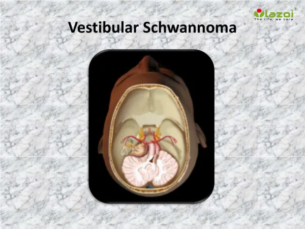
MONITORING OPERATIONS FOR VESTIBULAR SCHWANNOMA CHAPTER III
Monitoring of the facial nerve is a model for monitoring other cranial nerves
How to activate the motor system? • Electrical stimulation of motor nerves • Magnetic stimulation of motor nerves • Electrical stimulation of the motor cortex • Magnetic stimulation of the motor cortex
How to record the response? • Recording of electromyographic (EMG) potentials • Mechanical recordings of muscle contractions • Recording of motor nerve CAP
Recording muscle responses • Muscle relaxants cannot be used
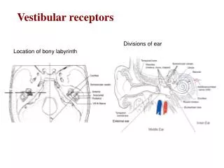
Monitoring of motor systems The facial nerve
Preservation of the facial nerve in operations for vestibular schwannoma • Identification regions of the tumor where there is no part of the facial nerve present • Identification of all parts of the facial nerve • Monitoring of mechanical induced facial nerve stimulation • Monitoring of injury induced facial nerve activation
Preservation of the facial nerve in vestibular schwannoma operations • Monopolar, constant voltage stimulation • Facial EMG made audible
Find the location of the facial nerve • Vary the strength of the stimulation to obtain less than maximal response • Note change in amplitude as stimulating electrode is moved • Increased amplitude of EMG means that the electrode was moved towards the facial nerve • Decreased amplitude of EMG means that the electrode is moved away from the facial nerve
Absence of mechanically induced EMG activity does not guarantee that injury has not occurred ! Always use electrical stimulation to verify the location of the facial nerve and its integrity
The likelihood of postoperative facial weakness • Increases with the number of occurrences spontaneous EMG • The duration of the activity is important
If the facial nerve is injured in the beginning of the operation (neurapraxia) It will not be possible to monitor the facial nerve during the remaining part of the operation
Use of partial muscle relaxation • Difficult to keep constant level of muscle relaxation • Prevent repetitive muscle contractions • Questionable whether partial muscle relaxation offers any protection of the patient from moving
Testing the function of the facial nerve For prediction of post operative facial function Always use electrical stimulation
Use of EMG for decision making regarding grafting? Electrophysiologic methods cannot distinguish between neurapraxia, axonotmesis or neurotmesis
Recording EMG from the masseter muscle (CNV) together with recording of facial EMG
Difference in EMG response to stimulation of the facial and the trigeminal nerves
The goals to reduce complications have been accomplished
This lady could have had facial palsy on one side after an operation for a vestibular schwannoma
Auditory neuromonitoring Recording of auditory evoked potentials in operations in the posterior fossa
Auditory monitoring for preservation of the function of the auditory nerve Recording of auditory evoked potentials in operations in the posterior fossa
Monitoring of ABR can detect manipulations of the brainstem before cardiovascular signs change
Use of ABR to detect manipulations of the brainstem in operations for large acoustic tumorsABR evoked from the contralateral ear has advantages over cardiovascular signs
Recording of ABR elicited from the contra-lateral ear monitor the brainstem
Recording of ABR elicited from the contra-lateral ear monitor the brainstem
Effect of brainstem manipulation AFTER BEFOREBLOOD PRESSURE Amplitude of peak V decreases SAME TIME AFTER BEFOREHEART RATE SAME TIME
Effect of brainstem manipulation SAME TIME BEFORE BLOOD PRESSURE Latency of peak V increases SAME TIME BEFORE HEART RATE
Waveform analysis of the ABR provides information about the anatomical location of an injury
NEURAL GENERATORS OF THE ABR: • Peak I: distal auditory nerve • Peak II: central auditory nerve • Peak III: mainly cochlear nucleus • Peak IV: unknown • Peak V: termination of • the lateral lemniscus in the contralateral inferior colliculus
Ipsilateral stimulation PEAK V Waveform analysis of the BAEP provides information about the anatomical location of an injury PEAK III
PEAK V Contralateral stimulation Waveform analysis of the BAEP provides information about the anatomical location of an injury PEAK III