Download

1 / 44
500 likes | 796 Views
Assessment of Cardiac Viability. Joyce Meng M.D. 8/15/2007. Viability-some definitions. The most practical definition of viability is “myocardium that demonstrates contractile dysfunction that shows functional improvement after revascularization” Stunning vs. hibernation

E N D
Assessment of Cardiac Viability Joyce Meng M.D. 8/15/2007
Viability-some definitions The most practical definition of viability is “myocardium that demonstrates contractile dysfunction that shows functional improvement after revascularization” • Stunning vs. hibernation • “Stunned”- transient post-ischemic dysfunction or myocardial contractile dysfunction in the presence of resting normal flow. • “Hibernating”- sustained abnormal contraction that can be attributed to chronic under-perfusion • Maybe a spectrum- there maybe a temporal progression from stunning, characterized by normal flow with reduced flow reserve to hibernation with reduced resting flow.
Pathophysiology • Pathologic features include: reduction in protein and mRNA synthesis, disorganization of the contractile elements (cytoskeletal proteins such as myosin, actin, desmin…etc by electron microscopy), and increase amount of extracellular matrix protein resulting in fibrosis • 38 patients with hibernating myocardium defined by thallium scintingraphy with re-injection and low-dose dobutamine echocardiography had cardiac biopsy during surgery to characterize the pathology of the hibernating myocardium circulation 97; 96 (9):2920-31
Viability • Different non-invasive methods that assess viability tests different facets which indicate that the “cell is alive”. • Delay enhancement with MRI shows scar. • Thallium and technecium uptake indicates intact cell membrane (thallium is a potassium analogs that relies on the Na/K ATPase for uptake, technecium uptake relies on intact mitochondrial membrane potential) • FDG-18 uptake indicates active metabolism. • Dobutamine echo, dobutamine MRI tests contractile reserve.
Viability • Likely that some characteristics (contractile reserve) while other more basic features (cell membrane integrity) persists. • At least partially explains the varying sensitivity and specificity of different techniques in predicting functional improvement.
Various endpoints indicating “viability” • Endpoints- improvement in • survival • symptoms • overall EF • Function of a particular segment (segments often defined in different ways in different studies) • Many studies focuses on the last objective while fewer studies address the more important issues.
Other problems with the data… • No study where treatment (medical vs revascularization) is randomized. Thus results are subjected to substantial referral bias. • Many studies are small and lack power. Hence, “overall” sensitivity and specificity are often derived from meta-analysis that group studies with different methodologies and length of follow up. • Few head to head comparison between methods.
Data supporting viability • Awaiting the result of the STICH trial. • Include patients with EF <=35% with anatomy suitable for revascularization. • Randomized to medical therapy, revascularization, or revascularization plus surgical scar excision. • Myocardial viability assessment by either radionuclide or echocardiographic techniques is not required but “strongly encouraged”. • Endpoints will include survival, cardiac mortality and morbidity, exercise capacity, LV size and function…etc.
Thallium- 201 • Potassium analog, myocardial uptake requires Na-K ATPase and is therefore indicative of cell membrane integrity and therefore, myocardial viability • Shows redistribution: initial uptake in areas that are well perfused, but slowly moves to areas that are alive but not well perfused. • Long physical 1/2 life (73 hours), limiting the overall amount that can be administered to 2-4 mCi. • Principle photo- peaks of mercury X-ray at 69 to 83kV (85-90%)
Thallium 201 protocols • Areas of ischemia by traditional stress- redistribution imaging is considered viable • injection of radiotracers during stress, acquire stress image within 10 minutes, wait 4 hours at rest, acquire a re-distribution image. Compare the two- if a defect present on the stress image resolved on the re-distribution image, the area is considered to be ischemic. • Persistent defects still show about 50% chance of improvement after revascularization
Thallium 201 protocols • If there is a persistent defect found, can use two methods to assess for viability. • Rest-redistribution • Stress- re-injection- delayed redistribution • Some variations in the two methods listed above.
Rest- redistribution inject 2-3 mCi of thallium at rest acquire the rest scan after 10 minutes wait for 4 hours acquire the re-distribution scan Stress- reinjection- redistribution Proceed with the usual protocol for a stress- redistribution scan If the defect is fixed, then reinject with 1 mCi of thallium Wait 24 hours later and acquire the delayed re-distribution image. Protocols
Indicators of viability • greater than 10% threshold increase in the tracer uptake compared to the previous image. • greater than 50% tracer uptake in a particular area. This is often done semi-quatitatively • comparing it to the brightest region • comparing this to a normal database • picture 2.jpg
Data • Meta-analysis: • Medline search from 1980 to 2000 • Included prospective study in patients with CAD who underwent revascularization whose results allowed assessment of sensitivity, specificity, PPV, and NVP • excluded patients with recent MI or UA (mostly within 1 month). • Major problems: • Most studies “segmental recovery” instead of overall improvement in EF, functional status, or survival • “segmental improvement’ is not standardized • Segments are not standardized
Data on rest-redistribution method • 22 mostly small studies (14-46 patients) with a total of 557 patients. • Some studies include consecutive patients • Patients are mostly male, the majority of whom have multivessel disease with a mean EF of approximately 25-40%, • functional recovery are assessed by Echo (15), some with RNV (5), or MRI (2). • Follow up varied from mean of 7 days to 12 months Bax JJ et al, Curr Probl Cardiol. 2001;26:142–186.
Data on rest-redistribution • 16 SPECT studies, 4 planar studies, 2 where camera was not named • 20 studies used conventional rest then 3-4 hour redistribution, 2 studies used rest then 24 hour delayed imaging • For analysis of results, 20 studies used a semiquantitative approach, 2 used visual estimates. • Viabiliy criteria- 2 studies used defect reversibility, 11 studies used Tl 201 activity cutoff level ranging from 50-75%, 9 studies used a combination of the two criteria. Bax JJ et al, Curr Probl Cardiol. 2001;26:142–186.
Results • Mean sensitivity 86% • Mean specificity 59% • PPV 69% • NNP 80% Bax JJ et al, Curr Probl Cardiol. 2001;26:142–186.
Data for stress-reinjection-delayed redistribution method • 11 (12-73 patients) with a total of 301 patients • Mostly older male with multivessl disease with mean EF of 31-50% • Function recovery were assessed by Echo (6) and RNV (6) • Follow up period also varied from 18 days to 6 months, although mostly in 3-6 month Bax JJ et al, Curr Probl Cardiol. 2001;26:142–186.
Data for stress-reinjection-delayed redistribution method • All but one studies used SPECT for image acquisition • 3 studies used a stress/rejection protocol (re-injection immediately after the stress test and acquiring the image 4 hours afterwards), while 8 used the stress-reinjection- delayed redistribution protocol • 8 studies used semiquantitative analysis while the remaining three used visual estimates • Viability criteria- 7 used a combination of defect reversibility and 50% maximal uptake while 4 used defect reversibility. Bax JJ et al, Curr Probl Cardiol. 2001;26:142–186.
Results • Mean sensitivity 88% • Mean specificity 50% • PPV 57% • NPP 83% Bax JJ et al, Curr Probl Cardiol. 2001;26:142–186.
Data on mortality • Several studies addressed this issue • 70 patients with multivessel disease and EF <40 underwent CABG • planar scans, rest- redistribution after 3 hours, thallium scan was divided into 15 segments, given score of 2 if uptake >75 and reversible, given score of 1 if uptake >50 and mildly fixed, and 0 if it is fixed. A viability index was generated by dividing score by the max score (30) • follow up 3 years • those with a viability score of >2/3 (medium) had better outcome- 6/33 death vs 17/37, p= 0.02 • The clinical profiles of the two groups are comparable at baseline. Pagley PR et al. Circulation. 1997;96:793–800
Technetium-99m sestamibi • Lipophilic cationic compound. Uptake across myocytes depend upon the presence of intact electrochemical gradients across the sacrolemmal and mitochondrial membranes. Thus it can be used as an agent to assess viability. Does not redistribute • Physical half life is 6 hours, can use higher dose (30mCi) because of the more favorable dosimetry profile. • Has a higher photon emission energy (140keV) and is well suited for gamma camera imaging.
Protocols for assessing viability • >50% uptake in the rest scan is a good predictor of viability • Nitrate-enhanced assessment • inject 10mg of isosorbide dinitrate diluted in 100ml saline infused for 20 minutes. As soon as the SBP dropped >20mmHg or SBP is <90, the tracer was injected. If none of these two criteria were met, the tracer was injected 15 minutes after the start of the infusion. Compare the imaged acquire during nitrate infusion with the image obtained at rest. The two scans were separated by 24 hours.
Data • 20 small studies (14-50 patients) with a total of 488 patients with CAD and LV dysfunction. 3 studies used 99mTc-tetrofosmin), 7 used nitrates • 17 used SPECT, 3 planer imaging • Used a combination of mostly echo and RNV to evaluate functional improvement. Several studies used MRI, gated SPECT, and contrast ventriculography • Viability defined as activity beyond a 50% maximal uptake or “redistribution with nirates” • Follow up as early as 20 days, but mostly in 3-6 months
Results • Mean sensitivity of 81% • Mean specificity of 66% • PPV of 71% • NPP 77% • When nitrate enhanced studies were included • mean sensitivity improved to 86%, specificity to 83%
prognosis • 105 patients with chronic CAD and LV dysfunction underwent nitrate-enhanced Tc-99-sestamibi imaging. They subsequently underwent medical treatment (group 1), complete revascularization (group 2A), or incomplete revascularization (group 2B) • Significantly worse event free survival curve was observed in group 1 and group 2B as compared to group 2A after approximately 2 years of mean follow up • Calculation of the Cox proportional hazard model reveals that the number of non-revascularized dysfunctional segment with viability on imaging is the most significant independent prognostic factor with a RR of 1.4 Sciagra R et al J Am Coll Cardiol. 2000;36:739–745
General principles of PET scans • Radionuclides used in PET studies are characterized by excess positive charge. • This unstable structure results in the emission of a positron (like an electron but with a positive charge) from the nucleus, thereby converting a proton to a neutron. • The positron travels a short distance (a few mm) until it encounters an electron. Annihilation ensues with the release of a photon pair traveling in opposite directions at the same time with a characteristic energy of 511 keV. The shorter the proton travels, the more “accurate” the tracer • When two opposing detectors sense the photons at the same time, its circuitry registers an event that occurred.This circumvent the use of collimators, increasing the sensitivity of the camera and allows for absolute count quantification
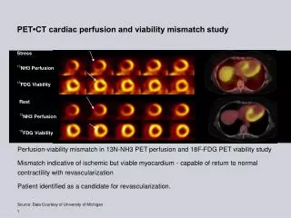
Indicators of viability • The most well-studied method compares the perfusion and metabolism of the heart. • Areas that are well perfused with metabolic activity are obviously viable • Areas with no perfusion but metabolic activity are also viable (mismatch) • Areas with no perfusion nor metabolic activity are not viable • Sometimes one notices area with good perfusion with little metabolic activity. Since it is perfused but shows poor contractile function it should be considered viable (result maybe consequence of poor glucose uptake due to diabetis…etc)
Most often used tracer includes N13 ammonia and Rb82 Both are reasonable perfusion tracers with high fractional extraction during 1st pass and linear relationship between net tissue extraction and blood flow up to 2.5x normal Neutral NH3 readily crosses the cell membrane and equilibrates with charged NH4, which gets trapped in the myocardium by being incorporated into glutamine. N13 has a physical ½ life 9.96 minutes 82Rb is a potassium analog with kinetic properties similar to 201 Tl with a physical ½ life of 76 seconds. The positron travels further prior to annihilation, thus there is more spatial uncertainty of the decaying nucleus. Perfusion tracers
18F- FDG • 18F-FDG is a glucose analog where one of the OH group is replaced by an 18F atom. • Initial uptake is comparable to glucose uptake • After phosphorylation, it remained trapped in the myocyte and cannot be further metabolized and therefore becomes a strong signal for imaging
Glucose metabolism in the myocardium • The myocardium typically uses 2/3 fatty acid oxidation and 1/3 glucose to meet its energy needs. • Uptake of glucose increases in the post-prandial state. • During ischemia, energy production is shifted from fatty acid oxidation to glucose which may contribute up to 70%of the total energy production
Glucose metabolism in the myocardium • Usually, the patient is asked to fast for 6 hours followed by administration of a statndard glucose load (25-100g) to stimulate natural insulin production • The myocardium of diabetic patients often have very poor glucose which results in suboptimal image quality • Can use hyperinsulinemic/euglycemic clamping • Often given a smaller oral load followed by small doses of IV insulin to achieve optimal blood glucose level prior to image acquisition
Data • In a group of heterogeneous 20 studies with total of 598 patients: • different perfusion tracers- some comparing to SPECT tracers • different criteria for viability- perfusion mismatch, normal perfusion, normalized FDG uptake, quantitative assessment of glc utilization • different metabolic state- s/p oral glucose loadng, using hyperinsulinemic euglycemic clamping, fasting, Bax JJ et al, Curr Probl Cardiol. 2001;26:142–186.
Results • For predicting segemental functional recovery the pooled data showed • Sensitivity of 93% • Specificity of 58% • mean PPV 71% • Mean NVP was 86% Bax JJ et al, Curr Probl Cardiol. 2001;26:142–186.
Prognosis • More data is available compared to Th-201 and Tc 99m sestamibi • Pooled 7 studies that studied long term prognosis of FDG/PET in a total of 619 patients demonstrates that event rate (death or MI) of • Viable/revascularized: 7% • Viable/medical treatment: 29% • Not viable/revascularized: 12% • Not viable/medical treatment: 12% • Analysis above suffers from non-uniformity: different definition of viability, different length of follow up…etc Bax JJ et al, Curr Probl Cardiol. 2001;26:142–186.
Comparison of Nuclear techniques with low dose dobutamine echocardiography
Head to head comparisons • 18 small studies (14-73 patients) with a total number of 563 patients directly compare viability assessment via a nuclear technique and low dose dobutamine echocardiography • 3 studies compared DE and FDG PET, the remaining studie compared DE with thallium imaging. • Pool results indicates that nuclear techniques have a higher sensitivity (88% vs 76%) and negative predictive value (80%vs 69%) for predicting segmental functional improvement, whereas DE has a higher specificity (53% vs 81%) and positive predictive value (84% vs 75%) Bax JJ et al, Curr Probl Cardiol. 2001;26:142–186.
Delayed enhancement • 50 patientswith chronic CAD and LV dysfunction undergoing revascularisation. • Recovery of function was assessed11 weeks post-revascularization. The likelihood of segmentalrecovery of function post-revascularization paralleled the extent. • Using a cutoff value of 25% transmurality of scar tissue, thesensitivity and specificity were 86% and 61% to predict improvementof function, respecitivly • Using 50%, the sensitivity and specificity were 97% and 44% • Using 75%,sensitivity and specificity would be 100% and 15%, respectively., Kim RJ et al, N Engl J Med 2000;343:1445–53
Conclusions • Viability assessment is probably useful, though data by severely limited by the lack of randomized trials heterogeneity of methods. • One of the largest meta-analysis- MEDLINE search of 24 studies reporting patient survival with a total of 3088 patients with viability assessment using thallium, FDG, and dobutamine echo. • Patients had EF of approximately 30% • Mean follow up was for about 2 years. • In patient with viability, revascularization was associated with an 80% reduction in annual mortality (16% vs 3.2%) compared with medical treatment. • In patient without viability, mortality was not improved by revascularization (7.7% vs 6.2%). J Am Coll Cardiol 2002 Apr 3;39(7):1151-8.
Viability assessment via various techniques • Assessment using Tl-201 and Tc 99m labeled agents appeared comparable with high sensitivity (mid-high 80%) and lower specificity (low-mid 60%). • F18 FDG PET appeared to be slightly more sensitive (sensitivity high 80-low 90%) but just as specific. It has more robust data regarding long-term prognosis. • Compared to low dose dobutamine echocardiogram, nuclear techniques have higher sensitivity (88% vs 76%) but lower specificity (53% vs 81%) • The sensitivity and specificity for segmental functional recovery depends on the extent of scar on delayed enhancement using MRI. Using a cut-off of 50% scar tissue, the sens/spec appear similar to FDG PET.