Download

1 / 37
380 likes | 430 Views
Learn about cardiac output measurement methods, preload and afterload factors, ventricular function curves, and determinants of pump function. Discover noninvasive and invasive techniques for evaluating cardiac performance.

E N D
Learning Objectives After reading this chapter you will be able to: Define cardiac output, cardiac index, stroke volume, and venous return Describe the following regarding cardiac output: Method of calculation Range of normal values Effect of sympathetic nervous stimulation
Learning Objectives (cont’d) Describe regarding the distribution of blood flow: Effect of metabolism and reduced oxygen availability on the regulation of blood flow through organs Percentage of total blood volume in venous system Effect of blood loss (hypovolemia) on circulatory function Basal distribution of blood flow to organs versus distribution during cardiac failure Effect of mechanical ventilation
Learning Objectives (cont’d) Explain the significance of the following indicators of cardiac output: Cardiac index, ejection fraction, stroke volume, end-diastolic volume, cardiac work, ventricular stroke work
Learning Objectives (cont’d) • Describe the following regarding preload: • Definition • Values used to measure preload of the left and right ventricles • Factors affecting • Clinical value of ventricular function curves • Effect of mechanical ventilation
Learning Objectives (cont’d) Describe the following for afterload: Definition Factors affecting Measurement Effect of vasodilators Calculation of systemic and pulmonary vascular resistance Effect of mechanical ventilation
Learning Objectives (cont’d) • Describe the following regarding contractility: • Definition • Factors affecting • Assessment
Describe the technique for obtaining cardiac output via the following invasive methods: Thermodilution Fick Pulse contour Doppler ultrasonic transducers Learning Objectives (cont’d)
Learning Objectives (cont’d) • Describe the noninvasive methods for evaluating cardiac performance: • Transthoracic electrical bioimpedance • Echocardiography • Radionuclide cardiac imaging • Partial CO2 rebreathing
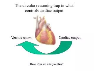
Cardiac Output (CO) The amount of blood pumped out of the left ventricle in 1 minute is the CO A product of stroke volume and heart rate Stroke volume: amount of blood ejected from the left ventricle with each contraction Normal stroke volume: from 60 to 130 ml Normal CO: from 4 to 8 L/min at rest
Venous Return Amount of blood returning to the right atria each minute is the venous return Normally venous return is the same as CO Venous return increases with peripheral vasodilation and decreases with vasoconstriction In a healthy heart, as venous return increases so does CO
Measures of Cardiac Output and Pump Function Cardiac index (CI) Determined by dividing the CO by body surface area Normal CI is 2.5 to 4.0 L/min/m2 CI measurement allows a standardized interpretation of the cardiac function
Measures of Cardiac Output and Pump Function (cont’d) Cardiac work A measurement of the energy spent ejecting blood from the ventricles against aortic and pulmonary artery pressures It correlates well with the amount of oxygen needed by the heart Normally cardiac work is much higher for the left ventricle
Measures of Cardiac Output and Pump Function (cont’d) Ventricular stroke work A measure of myocardial work per contraction It is the product of stroke volume times the pressure across the vascular bed Ventricular volume Estimated by measuring end-diastolic pressure
Measures of Cardiac Output and Pump Function (cont’d) • Ejection fraction • The fraction of end-diastolic volume ejected with each systole; normally 65% to 70%; drops with cardiac failure
Determinants of Pump Function 1. Heart rate (HR) Normally not a major factor in control of CO Extreme abnormalities can alter CO A low HR is normally compensated for by an increase in stroke volume (SV) A significantly elevated HR often causes SV to drop in people with heart disease when it reduces filling time
Determinants of Pump Function (cont’d) 2. Preload Created by end-diastolic volume The greater the stretch on the myocardium prior to contraction the greater the subsequent contraction will be When preload is too low, SV and CO will drop This occurs with hypovolemia Too much stretch on the heart can also reduce SV
Ventricular function curves A measure of heart output is placed on the vertical axis and end-diastolic volumes are placed on the horizontal axis Allows clinicians to see the changes in CO associated with various levels of preload In general, increases in preload will increase SV and CO until a physiologic end limit is reached Determinants of Pump Function (cont’d)
Ventricular compliance The stiffer the left ventricle the higher the preload needs to be to obtain an adequate SV Reduced ventricular compliance is caused by: Myocardial infarction Shock Pericardial effusions PEEP Positive inotropic drugs Determinants of Pump Function (cont’d)
Factors that affect venous return, preload, and CO Circulating blood volume Distribution of the blood volume Atrial contraction (adds 30% to subsequent ventricular SV) Determinants of Pump Function (cont’d)
Effect of mechanical ventilation Spontaneous inspiration lowers intrapleural pressures; improves venous return and CO Positive pressure breaths increase intrapleural pressures and reduce venous return and CO Degree of alteration in venous return with positive pressure breaths depends on the lung and chest wall compliance Venous return maximally reduced when lung compliance high, chest wall compliance is low Determinants of Pump Function (cont’d)
3. Afterload Two components: peripheral vascular resistance and tension in the ventricular wall Increases with ventricular wall distention and peripheral vasoconstriction As afterload increases, so does the oxygen demand of the heart Decreasing afterload with vasodilators may help improve SV but can cause BP to drop if the blood volume is low Determinants of Pump Function (cont’d)
Calculating SVR and PVR SVR is a measure of resistance to blood flow through the systemic circulation SVR increases with peripheral vasoconstriction and occurs with hypertension and use of vasoconstrictors PVR is a measure of pulmonary vascular resistance and increases with pulmonary vasoconstriction as seen in hypoxemia and acidosis Determinants of Pump Function (cont’d)
4. Contractility The final factor that determines pump function A measure of myocardial contraction strength Determined by: Amount of stretch on ventricle prior to contraction Inotropic state of the heart Reduced with hypoxia, acidosis, electrolyte abnormalities, and myocardial ischemia Determinants of Pump Function (cont’d)
Invasive Techniques Thermodilution Most common technique Requires placement of a pulmonary artery catheter and use of a computer A cold bolus of saline injected into a proximal port; temperature change over time measured by a thermometer at the tip of the PAC The degree of temperature change between the proximal port and the distal tip is a function of CO
Invasive Techniques (cont’d) Fick method Based on the fact that CO can be calculated if the oxygen consumption, the arterial oxygen content, and the mixed venous oxygen content are simultaneously measured Because this technique is very difficult to use regularly, it is not popular in the clinical setting
Continuous Cardiac Output Monitoring (CCO) Useful when patient is hemodynamically unstable and frequent measurements needed Indicator dilution techniques Arterial pulse contour analysis Can be done with invasive or noninvasive techniques After calibration CO is determined by the shape of the arterial pressure waveform
Transtracheal, Transesophageal, and Intravascular Ultrasound Doppler ultrasound can be placed on tubes or catheters to measure blood flow and CO The transtracheal ultrasound is attached to the end of an endotracheal tube and aimed at the descending aorta
Transtracheal, Transesophageal, and Intravascular Ultrasound (cont’d) Transesophageal ultrasound placed in the esophagus and aimed at descending aorta The intravascular ultrasound is placed on the distal end of a pulmonary artery catheter All three techniques require a bedside ultrasound technician and are expensive
Periodic Noninvasive Measurement of CO Echocardiography Provides periodic measurements of cardiac performance (ejection fraction, SV, CO) Can also diagnose shunts and other pathology Air trapping in the lung can make it difficult to image the heart
Radionuclide Cardiac Imaging Thallium-201 can be injected into the coronary arteries to look for areas of poor perfusion At the same time, the imaging can measure ventricular wall movement and ejection fraction
Blood Pressure Not a good indicator of blood flow A blood pressure cuff that examines the arterial pulse contour to estimate CO is available Pulse pressure is a reflection of stroke volume in many cases; narrow pulse pressures = low stroke volume
Summary Cardiac output monitoring a valuable part of hemodynamic assessment in critically ill patients Variety of invasive and noninvasive techniques are available The most popular invasive technique is the thermodilution RTs must be knowledgeable about the techniques used and the results obtained