Download

1 / 14
140 likes | 233 Views
Learn how to conduct a thorough cardiac assessment, including health history, inspection, palpation, and auscultation techniques. Understand normal vs. abnormal heart sounds and murmurs, and explore auscultation tips.

E N D
Comprehensive Cardiac Assessment • Health History • Inspection • Normal/abnormal • Palpation-4 landmark areas • Normal/abnormal • Technique • Auscultation
Auscultation • Normal • Rate • Rhythm • Regular • Irregular • Strength (intensity) • Extra Sounds
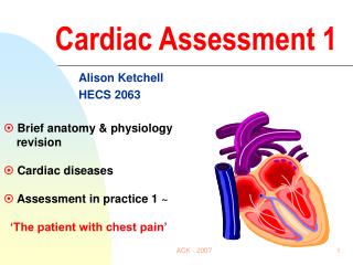
Anatomical Landmarks Heart Sounds • Aortic Area: 2nd intercostal space (ICS), right sternal border • Pulmonic Area: 2nd ICS, left sternal border • Tricuspid Area: 5th ICS, left sternal border • Mitral or Apical Area: 5th ICS, medial to the midclavicular line • AP TO MAN
S1: Systole • First heart sound • Closure of the AV valves – loudest at the tricuspid and mitral landmarks • “lub” • Dull, low pitched • Longer than S2 • Carotid pulse
S2: Diastole • “dub” • High pitch • Shorter • Semilunar valves close –loudest at the pulmonic or aortic valves landmarks • S1 and S2 within 1 second or less
S3 • Beginning of diastole • Position on L side • Mitral area • Bell • Low pitched • “Kentucky” • Too rapid of filling of the venticules • HF
S4 • Before S1 • Position on L side • Mitral area • Bell • Low pitched • “Tennessee” • Abnormal flow • Elderly, MI, HTN
Murmur • Turbulence of blood flow • Increased blood flow • Incomplete valve closure • Stenosis • Regurgitation • Blood flow through: • a dilated chamber • abnormal opening between chambers • Anywhere in the cycle • Pathology • Benign • Abnormal
Classification of Murmurs • Location • Intensity (I-VI) • Pitch • Quality • Timing • Position
Other Abnormal Heart Sounds • Pericardial Friction Rub • a scratching high pitched sound caused by friction between the pericardial and epicardial surfaces (pericarditis)
Cardiac Assessment Auscultation • Quiet environment • Consistent /Systematic Method– with bell and diaphragm • Listen for 1 full minute • Listen with stethoscope and feel for radial • Louder sounding valves at different locations • Positioning • Sitting up and leaning forward • Left lateral recumbent • Supine
Auscultation Tips • Diaphragm • high-pitched sounds such as S1, S2, murmurs, pericardial friction rubs. • Bell • low-pitched sounds such as S3, S4, and murmurs. • S3 and S4 best position is the Left lateral using the bell