Download
1 / 11
200 likes | 848 Views
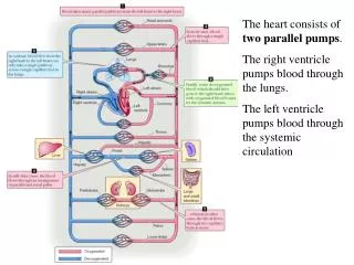
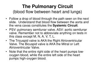
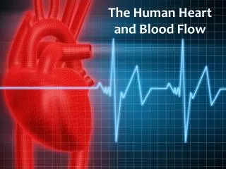
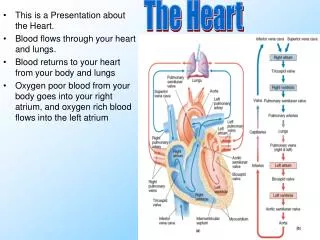
The Heart. This is a Presentation about the Heart. Blood flows through your heart and lungs. Blood returns to your heart from your body and lungs Oxygen poor blood from your body goes into your right atrium, and oxygen rich blood flows into the left atrium. How The Heart Works.

E N D
The Heart • This is a Presentation about the Heart. • Blood flows through your heart and lungs. • Blood returns to your heart from your body and lungs • Oxygen poor blood from your body goes into your right atrium, and oxygen rich blood flows into the left atrium
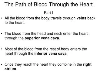
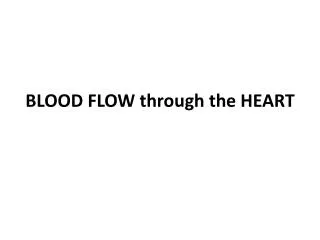
How The Heart Works. • The main function of the heart is to pump blood containing oxygen and nutrients to the body. • The blood is first pumped to the lungs to pick up oxygen and then to the rest of the body. Blood flows inside the heart, first going to the right atrium, then to the right ventricle, then to the lungs and finally to the left atrium and left ventricle.The heart beats almost like a clock, about 60 to 80 times per minute, whether you are asleep or awake. The heartbeat is regulated by a small "battery" located in the right atrium.
Blood Flow • Blood is pumped back out to your lungs and body. • Your right ventricle pumps blood out of your heart to your lungs, where the blood's oxygen supply is replenished. • At the same time, your left ventricle pumps blood — once again full of oxygen — out of your heart to your body.
Valves • There are four heart valves, mitral, aortic, pulmonary and tricuspid. • The tricuspid valve is between the right atrium and right ventricle. • The pulmonary or plutonic valve is between the right ventricle and the pulmonary artery. • The mitral valve is between the left atrium and left ventricle. • The aortic valve is between the left ventricle and the aorta. • Each valve has a set of flaps (also called leaflets or cusps). When working • properly, the heart valves open and close fully.
MRI of Heart. MRI stands for magnetic resonance imaging. It uses radiofrequency waves and a strong magnetic field rather than x-rays to provide remarkably clear and detailed pictures of internal organs and tissues. The procedure is valuable in diagnosing a broad range of conditions in all parts of the body, including heart and vascular disease, stroke, cancer and joint and musculoskeletal disorders. MRI is unique in that it can also create detailed images of blood vessels without the use of contrast material (although there is a trend toward the use of special non-iodinated
Nuclear medicine • Nuclear medicine is a healthcare specialty involving radioactive compounds to perform diagnostic imaging examinations that can lead to the effective treatment of many diseases. • Cardiac nuclear medicine tests are for people with unexplained chest pain or chest pain brought on by exercise to permit the early detection of heart disease. • The most common cardiac nuclear medicine procedure, is called myocardial perfusion scanning, which makes it possible to see the blood-flow patterns to the heart walls. The test is important for evaluating the presence and extent of suspected or known blockages.
Ultrasounds • An ultrasound test is a radiology technique that uses high frequency sound waves to produce images of the organs and structures of the body. The sound waves are sent through body tissues with a device called a transducer. • The transducer is placed on top of the skin, which has a gel applied to the surface. The sound waves that are sent by the transducer through the body are then reflected by internal structures as echoes. • These echoes return to the transducer and are transmitted electrically onto a monitor. The echo images are recorded on a plane film and can also be recorded on videotape. After the ultrasound, the gel is easily wiped off.
CAT Scans. • CAT stands for Computed Axial Tomography. • A CAT scan or CT scan is a painless test that uses x-ray images, taken from different angles, to create three-dimensional images of body structures. CAT scans use digital x-rays to produce their images on a computer screen
Septum • The septum is a part of your chest as well as a part of your nose • . The septum is the muscular wall separating the heart into the left and right sides. • The atrial septum is the wall separating the atria (the two upper chambers). • The ventricular septum is the wall separating the ventricles.