Download

1 / 62
620 likes | 727 Views
Explore the functions of the heart and lungs in maintaining optimal performance and health. Learn about the cardiovascular system's role in oxygen transport and its response to training. Discover the heart's structure, blood flow pathway, and cardiac cycle intricacies.

E N D
The Heart and Lungs at WorkChapter 6 Sport Books Publisher
Cardiovascular Fitness • Running is considered the most popular cardiovascular fitness program Sport Books Publisher
Learning Objectives 1. To develop an understanding of the organs and components of the human body that comprise the cardiovascular and respiratory systems. 2. To develop an understanding of physiological characteristics of the cardiovascular and respiratory systems and their functions to maintain health and optimal performance. 3. To develop an awareness of the measures that are used to evaluate and describe the various components of the cardiovascular and respiratory systems. 4. To develop an understanding of the effect of training on the cardiovascular and respiratory systems. Sport Books Publisher
The Primary Roles of the Cardiovascular System 1. to transport oxygen from the lungs to the tissues 2. to transport carbon dioxide from the tissues to the lungs 3. to transport nutrients from the digestive system to other areas in the body 4. to transport waste products from sites of production to sites of excretion. Sport Books Publisher
The Heart Structure • comprised of smooth muscle that serves to pump blood through the human body. • consists of four chambers: - two ventricles(left and right) pump blood through the body, - twoatria (left and right) receive blood from peripheral organs and pump blood into the ventricles • Left ventricle pumps blood through the entire body (are larger and with stronger muscle walls than the right ventricles) • Right ventricle pumps blood a short distance to the lungs Sport Books Publisher
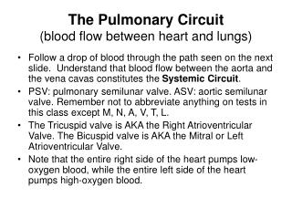
The Heart Pathway of blood flow: • The right atrium receives deoxygenated blood from the superior and inferior vena cava • The blood moves from the right atrium to the right ventricle and pumps it to the lungs • The left atrium receives the oxygenated blood from the lungs and pumps it to the left ventricle • The blood is now oxygen-rich and is transported to the entire body via the aorta Sport Books Publisher
Inferior vena cava Superior vena cava Deoxygenated Oxygenated The Heart Pathway of blood flow: RIGHT ATRIUM Tricuspid valve RIGHT VENTRICLE Veins Pulmonary semilunar valve Pulmonary arteries Capillaries Lungs Pulmonary veins Arteries LEFT ATRIUM Bicuspid valve LEFT VENTRICLE Aortic semilunar valve Aorta Sport Books Publisher
The Heart (b) Sodium-Potassium Pump (a) Chambers and Valves of the Heart Sport Books Publisher
The Heart Function • The heart contracts in a constant rhythm that may speed up or slow down depending on the need for blood (and oxygen) in the body. • The beating of the heart is governed by an automatic electrical impulse generated by the sinus node • The sinus node is a small bundle of nerve fibers that are found in the wall of the right atrium • The sinus node generates an electrical charge called an action potential. The action potential causes the muscle walls of the heart to contract. This action potential travels through the two atria and the two ventricles via the a-v node and the Purkinje fibres. • The atria contract before the ventricles contract, which allows for the blood to be quickly pumped into the ventricles from the atria Sport Books Publisher
(b) The mitral and tricuspid valves open, and the atria, squeezing into systole, force blood into the ventricles. (c) As the ventricle compartments fill with blood, they contract, thereby ejecting blood to the lungs and body. (d) The atria again relax and refill with blood. The Finely Tuned Cardiac Cycle (a) As the heart relaxes in diastole, both atria simultaneously fill with blood. Sport Books Publisher
The Heart Blood Pressure • This is an important measure of cardiac function. • There are two components to the measure of blood pressure: • Diastole -It is used to describe the pressure in the heart when the ventricles are relaxed and are being filled with blood. Indicator of peripheral blood pressure (the blood pressure in the body outside the heart). • Systole - It is the pressure in the ventricles when they are contracting and pushing blood out into the body. FYI: The normal range of pressure in the atria during diastole is about 80 mmHg, and during systole is about 120 mmHg. Sport Books Publisher
Measuring Blood Pressure • Doctor taking patient’s blood pressure Sport Books Publisher
The Heart Stroke Volume: • The amount of blood pumped out of the left ventricle each time the heart beats. • Measured in milliliters. • A typical stroke volume for a normal heart is about 70 milliliters of blood per beat. Cardiac Output: • The amount of blood that is pumped into the aorta each minute by the heart. • Cardiac output (ml/bpm) = stroke volume (ml) x heart rate (bpm) Sport Books Publisher
Measuring Heart Rate • Taking heart rate with fingers on wrist and neck (a) Feeling the carotid pulse (b) Feeling the radial pulse Sport Books Publisher
Maximum heart rate = 220 – age (years) The Heart Heart Rate • The number of times the heart beats in one minute, measured in beats per minute (bpm). • The contraction of the walls of the heart is commonly known as a heart beat. • The resting heart rate of an adult can range from 40 bpm in a highly trained athlete to 70 bpm in a normal person. • During intense exercise, the heart rate may increase to up to 200 bpm Sport Books Publisher
Circuitry of the Heart and Cardiovascular System Illustration of the entire cardiovascular system: heart, lungs, peripheral circulation Sport Books Publisher
The Heart The Peripheral Circulatory System • The peripheral circulatory system is comprised of the vessels that carry blood away from the heart to the muscles and organs (lungs, brain, stomach, intestines), and the vessels that return the blood to the heart. • All of the vessels of the body are made up of smooth muscle cells that allow them to contract or relax. • The contractile properties of smooth muscle enable the vessels of the peripheral circulatory system to regulate blood flow and alter the pattern of circulation throughout the body. Sport Books Publisher
Arteries Arterioles Capillaries The Heart The Peripheral Circulatory System • Vessels that carry blood away from the heart are called arteries. • Arteries branch into smaller and smaller vessels called arterioles. • The arterioles branch into even smaller vessels called capillaries. Sport Books Publisher
The Heart The Peripheral Circulatory System, Arteries cont’d • Capillaries: • allow for the exchange of oxygen and nutrients from the blood to muscles and organs • allow blood to pick up the waste products and carbon dioxide from metabolism Sport Books Publisher
The Heart The Peripheral Circulatory System, Veins • As the blood begins to return to the heart, the capillaries connect to form larger and larger vessels called venules. • The venules then merge into larger vessels that return blood to the heart called veins. Sport Books Publisher
The Heart The Peripheral Circulatory System, Veins continued • In comparison to arteries, veins have valves that open as blood returns to the heart, and valves that close as blood flows away from the heart. • Blood can be pushed through veins by smooth muscle that surrounds the veins, contraction of large muscles near the veins, or to a minor extent by the pumping action of the heart. Sport Books Publisher
The Skeletal Muscle Pump • blood flow towards the heart opens the valves • blood flow away from the heart closes the valves. Sport Books Publisher
The Heart Red Blood Cells • Also called erythrocytes • The primary function is to transport oxygen from the lungs to the tissues and remove carbon dioxide from the body. They are able to do this because of a substance called hemoglobin. • Other components of blood include white bloodcells and the clear fluid plasma. The percentage of the blood made up of red blood cells is called hematocrit (about 45%). Sport Books Publisher
The Red Blood Cell • Single red blood cell or erythrocyte Sport Books Publisher
The Heart Hemoglobin • A molecule made up of proteins and iron • Each molecule can bond to and transport four oxygen molecules. • The amount of oxygen that is carried by the blood is dependent upon the partial pressure of oxygen (PO2). • The difference in the amount of oxygen that is present in the blood as it leaves the lungs and the amount of oxygen that is present in the blood when it returns to the lungs is called the arterial-venous oxygen difference (a-v O2 difference), measured in ml of oxygen per litre of blood (ml O2 / l ) • If the a-v O2 difference increases, it means that the body is using more oxygen. • The typical a-v O2 difference at rest is about 4 to 5 ml O2 / l, while during exercise the a-v O2 difference can increase to 15 ml O2 / l. Sport Books Publisher
The Heart Hemoglobin • New red blood cells or reticulocytes are produced in the bone marrow • Erythropoietin (EPO),a circulating hormone, is the principal factor that stimulates red blood cell formation • EPO is secreted in response to low oxygen levels (when one goes to altitude) and also in response to exercise, thusincreasing the percentage of new red blood cells in the body • New red blood cells contain more hemoglobin than older red blood cells and thus can carry greater amounts of oxygen Sport Books Publisher
EPO Production • High altitude (low oxygen level) has an effect on EPO production which in turn generates a high production of red blood cells. Sport Books Publisher
CO2 + H2O H2CO3 Transport of Carbon Dioxide • CO2 is produced in the body as a by-product of metabolism • CO2 diffuses from the cells to the blood where it is transported to the lungs via one of three mechanisms: 1. A small percentage of the produced CO2 is dissolved in the blood plasma 2. CO2 bonds to the hemoglobin molecule 3. The primary mechanism whereby CO2 is transported through the body is via combining with water to form bicarbonate molecules that are then transported through the body. This happens according to the following reversible reaction Sport Books Publisher
Oxygen uptake • is the amount of oxygen that is consumed by the body due to aerobic metabolism • It is measured as the volume of oxygen that is consumed (VO2) in a given amount of time, usually a minute • Oxygen uptake increases in relation to the amount of energy that is required to perform an activity • (VO2max): a measure used to evaluate the maximal volume of oxygen that can be supplied to and consumed by the body Sport Books Publisher
Testing for Maximal Oxygen Uptake • Testing maximal aerobic power (VO2max) Sport Books Publisher
Oxygen Uptake • Changes in hematocrit (concentration of red blood cells in the blood) can also alter the oxygen uptake by increasing or decreasing the amount of oxygen that is supplied to working tissues. • The ability of the tissues to extract oxygen (a-vO2 difference) directly affects the oxygen uptake. • Increases in a-vO2 difference may arise due to an increased number of mitochondria in the muscles, or increased enzyme efficiency in working tissues Sport Books Publisher
Oxygen Uptake • Increased capillarization (number of capillaries in tissue) can affect the ability of the circulatory system to place red blood cells close to the tissues that are using the oxygen. • As a result, this increases the ability of those tissues to extract the required oxygen due to a shorter diffusion distance. Sport Books Publisher
VO2max = Cardiac Output x (a-vO2) difference Cardiovascular Anatomy Summary • The central component primarily concerns the effectiveness of the heart and the peripheral factors include; 1. the ability of the lungs to oxygenate the blood 2. the ability of the body to extract that oxygen. • Training can increase the maximal oxygen consumption of the human body. How this is accomplished will be presented in the next section. Sport Books Publisher
RESPIRATORY ANATOMY Sport Books Publisher
The primary role of the respiratory system is to: 1. deliver oxygenated air to blood 2. remove carbon dioxide from blood, a by-product of metabolism. The respiratory system includes: 1. the lungs 2. several passageways leading from outside to the lungs 3. muscles that move into and out of the lungs. Sport Books Publisher
The term respiration has several meanings: 1. ventilation (breathing) 2. gas exchange (occurs between the air and blood in the lungs and between the blood and other tissues of the body) 3. oxygen utilization by the tissues for cellular respiration. Sport Books Publisher
The Lungs • located within the thoracic cavity/chest. • the lungs are asymmetrical. The right lung is larger than the left lung because the heart takes up more space on the left side. • The air passages of the respiratory system are divided into two functional zones: 1. The conduction zone 2. The respiratory zone Sport Books Publisher
The Conduction Zone • the set of anatomical structures in which air passes before reaching the respiratory zone. • Air enters through the nose and or mouth, where it is filtered, humidified, and adjusted to body temperature in the trachea (windpipe). Sport Books Publisher
The Conduction Zone • The trachea branches into the right and left bronchithat enter the lung and continue to branch into smaller and smaller tubes called bronchiolesand finally the terminal bronchioles. • The whole system inside the lung looks similar to an upside-down tree that it is commonly called the “respiration tree”. Sport Books Publisher
The Respiration Zone • The region where gas exchange occurs. • The functional units of the lungs are the tiny air sacs, known as alveoli. • Alveoli are clustered in bunches like grapes, with a common opening into an alveolar duct called an alveolar sac. Sport Books Publisher
The Structure of the Respiratory System Sport Books Publisher
The Alveolus Sport Books Publisher
Ventilation and the Gas Exchange Oxygen In Carbon Dioxide Out Sport Books Publisher
Ventilation • Ventilation includes two phases,inspirationand expiration. Gas exchange between the blood and other tissues and oxygen utilization by the tissues are collectively known as internal respiration. Sport Books Publisher
Ventilation • Involves the movement of air into (inspiration) and out of (expiration) the lungs. • Changes in the size of the chest/thoracic cavity, and thus of the lungs, allow us to inhale and exhale air. • Lungs are normally light, soft and spongy to allow for expansion in the thoracic cavity. Sport Books Publisher
Ventilation • The muscles surrounding the thoracic cavity which result in size change include the: • Diaphragm • External Intercostal muscles (expiration) • Internal Intercostal muscles (inspiration) Sport Books Publisher
Ventilation • During inspiration, the thoracic cavity expands via muscle contractions causing the air pressure inside to be lowered. • The greater outside pressure causes a flow of air into the lungs. • During expiration, thoracic cavity shrinks via muscle relaxation • The greater outside presure causes a flow of air out of the lungs Sport Books Publisher
Gas Exchange in the Lungs • Gas exchange between the air and blood in the lungs occurs at the alveoli. • Each bubble-like alveolus is surrounded a vast network of pulmonary capillaries. • The atmospheric air which has made its way into each alveolus is rich in oxygen. Sport Books Publisher
Gas Exchange in the Lungs • The blood in the pulmonary capillaries is loaded with the waste product of carbon dioxide. This difference in concentration of C02 and O2 gases sets up ideal conditions for gas diffusion. • Diffusion is the movement of molecules (in this case, gases) from a higher concentration to a lower concentration • Therefore, oxygen diffuses through the alveolar membrane into deoxygenated pulmonary capillaries. Sport Books Publisher
Gas Exchange in the Lungs • Carbon dioxide diffuses in the opposite direction, from the carbon dioxide rich pulmonary blood into the alveoli • The oxygenated blood follows the pulmonary circulation to reach the heart (right ventricle) and is distributed through systemic circulation. • Carbon dioxide is exhaled out. Sport Books Publisher