Download
1 / 30
350 likes | 695 Views
Benign disorders of WBCs. By/ Mr. Waqqas Elaas; M.Sc; MLT. References. For theory : Essential Haematology, John Wiley & Sons Ltd ,6th Edition, Victor Hoffbrand . For practical : Practical Haematology, Churchill Livingstone, Eighth edition, John V. Dacie, S. M. Lewis,

E N D
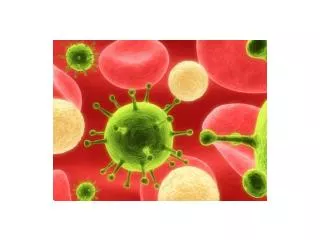
Benign disorders of WBCs By/ Mr. Waqqas Elaas; M.Sc; MLT
References • For theory : Essential Haematology, John Wiley & Sons Ltd ,6th Edition,Victor Hoffbrand. • For practical : Practical Haematology, Churchill Livingstone, Eighth edition, John V. Dacie, S. M. Lewis, • Internet site(s): • http://www.essentialhaematology6.com/default.asp = MCQs • http://www.hematologyatlas.com/ • http://pathy.med.nagoya-u.ac.jp/atlas/doc/atlas.html • Final Theoretical exam : 40 • Final Practical exam : 20 (including written questions) • 1st Periodic exam : 10 theory, 5 Practical • 2nd Periodic exam : 10 theory, 5 Practical • Homework and class activities : 5 Theory, 5 Practical • Total : 100 Marks
Objectives • To differentiate between the qualitative & quantitative WBCs benign disorders. • To understand the etiology and pathology of reactive changes in the number and morphology of granulocytes. • To understand the etiology and pathology of reactive changes in the number and morphology of lymphocytes and monocytes. • To know the definition & causes of Infectious Mononucleosis. • To know the definition & causes of Leukemoid reactions. • To be able to differentiate between Eosinophilia & Hypereosinophilic syndromes.
Leucocytes (WBCs) Phagocytes Immunocytes (Granulocytes) (A granulocytes) Neutrophils Lymphocytes Eosinophils small & Large Basophils B & T Lymphocytes Monocytes* *sometimes Monocytes are considered as A granulocytes
LEUCOCYTES BENIGN DISORDERS • Quantitative • Change in number • Terminology • Cytosis / philia • Increase in number • Cytopenia/penia • Decrease in number • Qualitative • Morphologic changes • Functional changes
LEUCOCYTES BENIGN DISORDERSQuantitative changes Relative & Absolute values To make an accurate assessment, consider both relative and absolute values. For example a relative value of 70% neutrophils may seem within normal limits; however, if the total WBC is 20,000, the absolute value (70% of 20,000) would be an abnormally high count of 14,000.
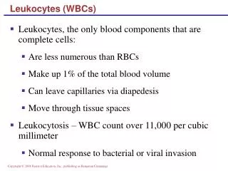
LEUCOCYTOSIS • Definition Raised TWBC above 11.0 x 10⁹/L in adults, due to elevation of any of a single lineage. • Note: elevation of the minor cell populations can occur without a rise in the total white cell count. • Normal reference range (adults) • 4.5 -- 11.0 x 10⁹/L
LEUCOPENIA • Definition TWBC lower than 4.5 x 10⁹/L in adults • Leucopenia may affect one or more lineages and it is possible to be severely neutropenic or lymphopenic without a reduction in total white cell count.
(contd.) • Granulocytosis Increase in the count of all or one of the granulocytic component: • Neutrophils • Basophils • Eosinophils
NEUTROPHILIA • Definition • Increase in the number of neutrophils and / or its precursors • In adults count >7.5 x 10⁹/L but the counts are age dependent • Increase may results from alteration in the normal steady state of • Production • Transit • Migration • Destruction
NEUTROPHILIA (contd.) • Causes of Neutrophilia • Infection • Bacterial • Inflammatory conditions • Autoimmune disorders • Gout • Neoplasia • Metabolic conditions • Uraemia • Acidosis • Haemorhage • Corticosteroids • Marrow infiltration/fibrosis • Myeloproliferative disorders
Leukemoid reactions • Excessive reactive leucocytosis. • Applied to chronic Neutrophilia with marked leucocytosis (>20 x 109/L) • The usual feature is the shift to the left of myeloid cells • Causes include • Infections • Marrow infiltration • Systemic disease (e.g.: Acute liver failure) • (Left shift : indicates that the neutrophils present in the blood are at a slightly earlier stage of maturation than usual. The Band and the stages before. This is often seen in acute infections). • (Right shift : an increase in the percentage of multilobed neutrophils).
NEUTROPENIA • Neutropenia is an absolute reduction in the number of circulating neutrophils • Mild (1- 1.5 x 10⁹/L) • Moderate (0.5 – 1 x 10⁹/L) • Severe (<0.5 x 10⁹/L) • Symptoms are rare with the neutrophil count above 1 x 10⁹/L • Bacterial infections are the commonest. • Fungal, viral and parasitic infection are relatively uncommon.
(NEUTROPENIA) contd. • Causes of Neutropenia • Racial • Congenital • Marrow aplasia • Marrow infiltration • Megaloblastic anemia • Acute infections • Typhoid, Miliary TB, viral hepatitis • Drugs • Irradiation exposure • Immune disorders • HIV • SLE • Neonatal isoimmune and autoimmune neutropenia • Hyperslplenism
(EOSINOPHILIA) • Increase in the eosinophil count must prompt for further investigation (>0.6 x 10⁹/L) • The causes of eosinophilia can be considered under following headings • Allergy • Atopic, drug sensitivity and pulmonary eosinophilia • Infection • Parasites, recovery from infections • Malignancy • Hodgkin’s disease, NHL and myeloproliferative disorders • Drugs • Skin disorders • Gastrointestinal disorders • Hypereosinophilic syndrome
(EOSINOPHILIA) Contd. • Hypereosinophilic syndrome • Criteria of diagnosis • Peripheral blood eosinophil >1.5 x 10⁹/L • Persistence of counts more than 6 months • End organ damage • Absence of any obvious cause for eosinophilia • Organ most commonly involved • Heart • Lung • Skin • Neurological
(MONOCYTOSIS) • Absolute monocyte count is age dependent • Count rarely exceeds >1.0 x 10⁹/L • Have no marrow reserves • Causes of monocytosis can be grouped as • Infections • Chronic infection (TB, typhoid fever, infective endocarditis) • Recovery from acute infection • Malignant disease • MDS, AML, HD, NHL • Connective tissue disorders • Ulcerative colitis, Sarcoidosis, Crohn’s disease • Post splenectomy
(BASOPHILIA) • Basophils are least common of the granulocytes • Reference range for adult is 0 – 0.2 x 10⁹/L • Most commonly associated with hypersensitivity reactions to drugs or food • Inflammatory conditions e.g RA, ulcerative colitis are also sometime associated with basophilia • Myeloproliferative disorders • Chronic myeloid leukemia
(LYMPHOCYTOSIS) • The blood contain only few percent of total body lymphocytes • The most consistent variation is seen with age • Alteration of lymphocyte counts can result from • The redistribution of lymphocytes • Absolute increase of lymphocyte number • Loss of lymphocytes • Combination of these
(LYMPHOCYTOSIS) • Non-malignant causes of lymphocytosis • Infections • Viral infections • Infectious mononucleosis • CMV • Rubella, hepatitis, adenoviruses, chicken pox,dengue • Bacterial infections • Pertussis • Healing TB, typhoid fever • Protozoal infections • Toxoplasmosis • Allergic drug reactions • Hyperthyroidism • Splenectomy • Serum sickness
(LYMPHOCYTOSIS) • Infectious Mononucleosis • Epstein-Barr virus • Saliva from infected person is the main contagion • Virus infect epithelial cells and B cells • Infection in children under the age of 10 does not cause illness and result in life long immunity • Clinical features • Fever, malaise, fatigue, sore throat, splenomegaly • Blood picture shows leucocytosis ( 10 – 20 x 109/L) due to absolute increase in lymphocytes • Diagnosis is by serological tests • There is no specific treatment
Qualitative changes (MORPHOLOGY) Congenital acquired Pelger-Huet anomaly Toxic granulation Neutrophil hyper-segmentation Dohle bodies May-Hegglin anomaly Pelger cells Alder’s anomaly Hypersegmented neutrophils Chediak-Higashi syndrome
LEUCOCYTES BENIGN DISORDERSQualitative changes (MORPHOLOGY) • Congenital • Pelger-Huet anomaly • Bilobed and occasional unsegmented neutrophils • Autosomal recessive disorder
LEUCOCYTES BENIGN DISORDERSQualitative changes (MORPHOLOGY) contd. • Neutrophil hyper-segmentation • Neutrophil function is essentially normal • May-Hegglin anomaly • Neutrophils contain basophilic inclusions of RNA • Occasionally there is associated leucopenia, Thrombocytopenia and giant platelet are frequent
LEUCOCYTES BENIGN DISORDERSQualitative changes (MORPHOLOGY) contd. • Alder’s anomaly • Granulocytes, monocytes and lymphocytes contain granules which stain purple with Romanowsky stain • Granules contain mucopolysaccharides
LEUCOCYTES BENIGN DISORDERSQualitative changes (MORPHOLOGY) contd. • Chediak-Higashi syndrome • Giant granules in granulocytes, monocytes and lymphocytes • Depressed migration and degranulation • Recurrent pyogenic infections • Lymphoproliferative syndrome may develop • Treatment is BMT
LEUCOCYTES BENIGN DISORDERSQualitative changes (MORPHOLOGY) contd. • Acquired • Toxic granulation • Dohle bodies • Pelger cells • Hypersegmented neutrophils
Homework (1) Case : A 20-year-old student presented with a 7-day history of fever, sore throat, lethargy and tender enlarged glands in the neck. Physical examination reveals fever, mild jaundice, inflamed pharyngeal mucosa and cervical adenopathy. Blood results Hb; 12.5 g/dl, wbc 18.0x10⁹/l , differential 30% neutrophils 40% lymphocytes 30% abnormal lymphocytes. Platelets 100 x109/l. Throat swab: No bacterial growth HIV test negative • Does the student has Neutrophilia OR Lymphocytosis? • Explain your answer in Q1 • What is the probable diagnosis? (2) Design a table containing the 5 types of leucocytes with their normal ranges in adults.