Download
1 / 33
450 likes | 1.9k Views
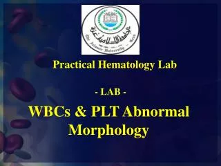
Practical Hematology Lab. - LAB -. WBCs & PLT Abnormal Morphology. Platelet Satellitism. Morphology Platelets clumped around neutrophils . Found in EDTA in vitro induced change of no clinical significance except false low platelet count (in vitro). Giant Platelets. Morphology

E N D
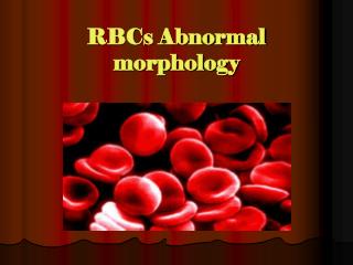
Practical Hematology Lab - LAB - WBCs & PLT Abnormal Morphology
Platelet Satellitism MorphologyPlatelets clumped around neutrophils. Found inEDTA in vitro induced change of no clinical significance except false low platelet count (in vitro).
Giant Platelets Morphology Platelet larger than a normal red cell. Found in • Increased platelet turnover • Myeloproliferative disorders • Myelodysplasticdisorders
Large Platelets MorphologyLarge platelets - larger than one third but less than the size of a red cell. Found in • Increased turnover of platelets • Myeloproliferative disorders • Myelodysplastic disorders • May Hegglinanomaly • Grey platelet syndrome • Bernard Soulier
Micro Clots MorphologyFibrin strands, platelets and white cells (in this case - neutrophils) clumped together. Found inIn vitro artifact caused by poor venesectiontechnique leads to false low counts - can influence white cell, red cell and platelet counts
Platelet Clumping MorphologySmall clumps of platelets. Found in • In vitro artifact caused by EDTA or cold and leads to false low platelet count. • Difficult venesection
Wiskott Aldrich Syndrome MorphologySmall platelets. Found in • Wiskott Aldrich syndrome
Grey Platelet Syndrome MorphologyPlatelets appear degranulated. Found in • Grey platelet syndrome • Discharge of platelet granules in vivo (cardiopulmonary bypass, hairy cell leukemia) • Discharge of platelet granules in vitro (poor venesection technique)
Drumstick MorphologyDrumstick shaped nuclear appendage. ± 1,5 µm in diameter and attached to the nucleus by a filament. Inactive X chromosome of the female. Found in • Neutrophils of females • Males with Klinefelter syndrome
Sessile Nodule MorphologyInactive X chromosome found as nodule on neutrophils of females. Found in • Neutrophils of females
Hypersegmentation or right shift of neutrophil nuclei MorphologyAverage lobe count increased OR increased % of neutrophils with 5 - 6 lobes OR > 3% neutrophils with 5 lobes or more.Found in • Megaloblasticanaemia • Iron deficiency • Chronic infection • Liver disease • Uraemia
Ring shaped nuclei MorphologyNucleus ring or doughnut shaped. Found in • Acute myeloid leukemia • Chronic granulocytic leukaemia • Megaloblasticanaemia • MDS
Detached Nuclear Fragments MorphologyDetached nuclear material in cytoplasm. Found in • Dysgranulopoiesis • Patients on anti cancer chemotherapy • HIV
Toxic Granulation MorphologyIncreased granulation. Granulation more basophilic and larger than normal. Found in • Severe bacterial infection. • Non specific finding - seen in tissue damage of various types. • Normal pregnancy. • Therapy with cytokines
Hypogranulation MorphologyReduced granulation in neutrophil cytoplasm. Found in • Myelodysplasticsyndromes
DohleBodies MorphologySmall pale blue cytoplasmic inclusions, often in the periphery of the cell. Consist of ribosomes and endoplasmic reticulum Found in • Infective and inflammatory states. • Severe burns • Tuberculosis • Post chemotherapy • Pregnancy • May-HegglingAnomaly
Russell bodies Morphology are eosinophilic, large, homogenous immunoglobulin-containing inclusions usually. Found in aplasma cell undergoing excessive synthesis of immunoglobulin; the Russell body is characteristic of the distended endoplasmic reticulum This is one cell variation found in multiple myeloma.
PhagocytosedParasites MorphologyMalaria - Plasmodium falciparum Found in • Severe malaria infection
PhagocytosedPlatelet MorphologyPlatelet in vacuole in neutrophil cytoplasm Found in • Infection
Phagocytosed Red blood cell MorphologyRed cell in vacuole in cytoplasm of neutrophil Found in • Infection • Auto immune haemolyticanaemia • Incompatible blood transfusion
Auer Rods MorphologySmall azurophil rods in the cytoplasm of myeloblasts and promyelocytes. Sometimes found in mature neutrophils. Found in • Acute myeloblasticleukemia. • Myelodysplastic syndromes.
Macro Neutrophils MorphologyTwice the size of a normal neutrophil with tetraploid DNA content. Found in • Occasionally in the blood of healthy subjects. • Inherited • Administration of G-CSF • Megaloblasticanaemia • Chronic infection
Necrobiotic / Apoptotic neutrophil MorphologyDense homogenous nuclei (pyknotic) Found in • Occasionally in healthy subjectsIn vitro artifact. • AML
Shift To The Left MorphologyPresence of precursor of granulocytes in the peripheral blood Found in • Normal in pregnancy or neonate. • Infections. • Bone marrow fibrosis. • Bone marrow infiltration by malignancies.
Pseudo PelgerHuet Anomaly MorphologyBilobed neutrophils with more condensed chromatin Found in • Inherited Myelodysplasticsyndromes. • Idiopathic myelofibrosis. • Chronic granulocytic leukemia. • Therapy with colchicine, ibuprofen. • Infectious mononucleosis, malaria, myxedema. • CLL
Neutrophil aggregation MorphologySmall clumps of neutrophils. Happens in vitro if EDTA anticoagulated blood is allowed to stand. May lead to incorrect WBC. Found in • In vitro finding. • Infectious mononucleosis. • Bacterial infections. • Auto immune disease.
Atypical Lymphocytes MorphologyPleomorphic. Large with diameter of 15 - 30 µm. Abundant, strongly basophilic cytoplasm. Basophilia may be confined to the cytoplasmic margins. Found in • Viral infections - EBV, CMV, Hep A, Measles. • Bacterial infections - brucella, tuberculosis. • Protozoa – malaria. • Immunization. • SLE.
PlasmacytoidLymphocyte MorphologyLymphocyte with basophilic cytoplasm and eccentric nucleus. Found in • Reactive phenomenon
Mott cell MorphologyPlasmacytoid lymphocyte with globular inclusions composed of immunoglobulin. Found in • Reactive changes in peripheral blood.
Large Granular Lymphocyte MorphologySmall eosinophilic granules in the cytoplasm of large lymphocytes. Found in • Natural killer cells. • Lymphokineactivated T cells.
Monocyte Vacuolization Morphology Vacuoles in the cytoplasm of monocytes. Found in • Infections
Chediak-Steinbrink-Higashi Anomaly Chediak-Higashi syndrome (CHS) is a rare autosomal recessive disorder characterized by recurrent pyogenic infections, partial oculocutaneousalbinism, progressive neurologic abnormalities, mild coagulation defects.