
Nervous System
Nervous System. The Brains of the Operation. Functions. Three major functions: 1 . Receive sensory input Gather info by monitoring changes or stimuli from inside & outside body 2. Integration of input Process & interpret sensory input & determine action 3. Motor output

Nervous System
E N D
Presentation Transcript
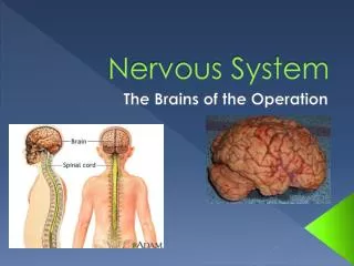
Nervous System The Brains of the Operation
Functions • Three major functions: • 1. Receive sensory input • Gather info by monitoring changes or stimuli from inside & outside body • 2. Integration of input • Process & interpret sensory input & determine action • 3. Motor output • Carry out response decided by integration usually by muscles (movement) or glands (secretion)
Organization • Two major parts of nervous system: • Central nervous system (CNS) • Brain • Spinal cord • Peripheral nervous system (PNS) • All nerves (spinal & cranial) outside of CNS • Two components: • Sensory (afferent) division • Info going TOWARD CNS • Motor (efferent) division • Impulses EXIT from CNS
Two subdivisions of motor (efferent): • Somatic nervous system – voluntary • Conscious control of skeletal muscles • Autonomic nervous system - involuntary • Controls smooth/cardiac muscle & glands
Two subdivisions of autonomic that often bring about opposite effects: • Parasympathetic – stimulate rest & digest activities • Ex: stimulate flow of saliva • Sympathetic – stimulate flight or fight activities • Ex: inhibit flow of saliva
Structure of Nervous Tissue • Classified into two types of cells: • Neurons – transmit nerve impulses; no cell division (amitotic) • Support cells (called neuroglia or glia) that can not transmit nerve impulses; cell division (mitotic) • Astrocytes • Microglia • Ependymal cells • Oligodendrocytes • Satellite cells • Schwann cells Central nervous system (CNS) Peripheral nervous system (PNS)
Neurons • Also known as nerve cells • Structure allows them to receive & transmit messages or impulses • All have same basic structures • Cell body – usual cell organelles including nucleusexcept no centrioles (no mitosis) • Processes/fibers – arms that extend to/from body • To body = dendrites (1-100s) • From body = axon (only 1)
Must know cell parts: • Soma - cell body • Nucleus – metabolic center of cell • Dendrite(s) – one or more processes that conducts impulses TOWARD cell body • Axon hillock – where cell branches out to axon • Axon – process that conducts impulses AWAY from cell body • Myelin – whitish, fatty substance found on long axons in CNS; speeds up transmission rate • Schwann cells – cells that myelinate axons in PNS • Myelin sheath – membranes wrapped around myelin • Nodes of Ranvier– gaps in between Schwann cells • Axon terminal – branched end of axon in which neurotransmitters are stored in vesicles
Large concentrations of cell bodies in CNS are in clusters & called nuclei • Small concentrations are called ganglia (pl) or ganglion (sing) – found in CNS & PNS • Bundles of nerve fibers in CNS called tracts, but in PNS are called nerves • Myelinated nerve fibers/tracts in CNS called white matter • Unmyelinated fibers and cell bodies called gray matter
Three types of neurons based on function or direction of nerve impulse: • Sensory (afferent) neurons • Carry impulses from sensory receptors TOWARD CNS • Nerve endings – pain & temperature receptors • Meissner’s corpuscle – touch receptor • Pacinian corpuscle – deep pressure receptor • Proprioceptors – stretch or tension in tendons & muscles • Motor (efferent) neurons • Carry impulses FROM CNS to organs or muscles • Association neurons/interneurons • Connect motor & sensory neurons
Functional Neuron Types (afferent) (efferent)
Three types of neurons based on structure or how many processes extend from body • Unipolar • Single, very short process from cell body • Immediately breaks into peripheral & central axon • Unique: dendrites at peripheral end, so axon conducts impulses away and TO cell body • Bipolar • One axon, one dendrite • Very rare, only seen sense organs (eye & nose) • Act as receptor cells • Multipolar • Several processes extend from cell body • All motor neurons are multipolar so they are most common
2. Support Cells2a. Astrocytes • Star-shaped • Account for ~ 50% of neural tissue • Form living barrier between capillaries & neurons therefore make exchanges between them • Help protect neurons from harmful substances • Pick up extra ions • Recapture released neurotransmitters
2b. Microglia • Spiderlike phagocytes (cell eaters) • Dispose of debris like dead brain cells & bacteria
2c. Ependymal Cells • Covered with cilia • Line cavity of brain & spinal cord • Beating of cilia help circulate cerebrospinal fluid that fills brain & spinal cord cavity • Forms protective cushion around CNS
2d. Oligodendrocyte • Wrap flat extensions tightly around nerve fibers • Produces fatty insulating covering of axons called myelin sheaths in CNS
2e. Schwann Cells • Form myelin around axons in PNS
2f. Satellite Cells • Protective, cushioning cell body in PNS
D. Nerve Impulse • neuron has two distinct properties that differentiate it from any other cell in human body: • Irritability - ability to respond to stimuli & convert it to a nerve impulse • conductivity - ability to transmit an impulse to other neurons, muscles, or glands • Most CNS neurons receive chemical stimulus at plasma membrane (everywhere on neuron), transmits it as electrical signal along axon, & ends as chemical signal at axon terminals • Most PNS neurons (sensory organs) receive stimulus as light (eyes), sound waves (ears), pressure (touch), or chemicals (taste & smell)
plasma membrane is where nerve impulse begins • Plasma membrane at rest is polarized • fewer positive ions (K+) are inside cell than positive ions (Na+) outside cell • More negative (Cl-) ions inside cell than outside - Na+ Na+ Na+ - Na+ Na+ Na+ Na+ Na+ Na+ Na+ Na+ - - - - - - - - - - - K+ K+ K+ K+ - K+ - K+ - K+ - - - - - K+ - K+ - - - Na+ Na+ Na+ Na+ - Na+ Na+ - Na+ Na+ Na+ Na+
a stimulus depolarizes neuron’s membrane by opening up Na+ gates on membrane, allowing Na+ inside • initial exchange of ions is a local depolarization • Inside is more + than outside • Depolarization starts an action potentialin entire neuron
Once action potential (nerve impulse) starts, it’s propagated over entire axon (all or nothing principle) • K+ ions rush out of the neuron after Na+ ions rush in, which repolarizes the membrane • Na+/K+pump on membrane restores original configuration by shoving Na+ back out and allowing K+ back in • requires ATP
Action Potential Analogy Polar = opposite ends INSIDERS outsiders outsiders outsiders outsiders INSIDERS INSIDERS INSIDERS INSIDERS INSIDERS INSIDERS outsiders outsiders INSIDERS outsiders outsiders Polarized (rest) Depolarized (action potential) Repolarized Polarized
Impulse travels faster when fibers have a myelin sheath • Once electrical action potential reaches axon terminals, excites vesicles containing neurotransmitters • Vesicles move toward axon terminal membrane & releases neurotransmitter into synaptic cleft • Neurons NEVER touch other neurons • Neurotransmitters bind to receptors on neighboring neuron’s dendrites • New action potential will start on THAT one
The human body uses 50 different neurotransmitters depending on the need • Neurotransmitters either excite or inhibit neurons • Many drugs act to mimic the effect of neurotransmitters on the brain
E. Reflexes • Much communication between neurons on everyday basis is done via reflexes • Reflex: rapid, predictable, involuntary responses to stimuli • Reflex always occurs in same manner using same neural pathways of both CNS & PNS so they are called reflex arcs
Two types of reflexes: • Somatic: stimulates skeletal muscles • Ex: pull hand away from hot object, blinking when air burst aimed at eyes • Autonomic: regulate smooth & cardiac muscles, & glands • Ex: secretion of saliva, change in pupil size
Reflex arcs have at least 5 elements involved in same arc or pattern: • Sensory receptor – react to stimulus • Sensory neuron – connect receptor & CNS • Integration center – connect neurons • Motor neuron – connect CNS & effector • Effector organ – muscle/gland to be stimulated
patellar (knee-jerk) reflex is simplest type of reflex – two neurons involved • Withdrawal reflex (remove from painful stimulus) is more complicated – three neurons involved utilizing association neuron
F. Brain Structure & Functions • Average adult brain weighs 3 lbs • Divided into 4 regions: • Cerebrum – largest region, broken into left & right hemispheres • Diencephalon – interbrain atop brain stem • Brain stem – stalk on which brain sits, connects to spinal cord • Cerebellum – bulbous projection at occipital region, broken into two hemispheres
1. Cerebrum • Made of two hemispheres together called cerebrum • Encloses other three parts of brain • Entire surface made of peaks and valleys • Gyrus (gyri) – peaks of ridges • Sulcus (sulci) – shallow valleys • Fissures – deep grooves separating large regions
Function of cerebrum is vast • speech, memory, logical & emotional response, consciousness, interpretation of sensation, voluntary movement • Sulci & fissures divide cerebrum into lobes (named after cranial bones) • Parietal lobe • Frontal lobe • Temporal lobe • Occipital lobe
Parietal Lobe • Somatic sensory area located just posterior to central sulcus receives & interprets impulses from body’s sensory receptors (NOT special senses) • Pain, cold, light touch
Spatial map depicting region on body where senses come from and how much brain power is devoted to them is called sensory homunculus Model depiction showing areas of body given more brain “power” than others
Sensory pathways are crossed pathways, meaning left side of brain receives impulses from right side of body & vice versa • Itch on right hand interpreted on left side of somatic sensory area.
Occipital lobe • Visual area located in posterior part • Visual cortex – “sees” • Visual association area – ties object to memories, knowledge • Temporal lobe • Auditory area bordering lateral sulcus • Auditory cortex – “hears” • Auditory association area – ties sound to object or knowledge • Olfactory (smell) area deep inside
Frontal lobe • Contains primary motor area, just anterior to central sulcus, which allows us control of skeletal muscles • Spatial map region called motor homunculus • Broca’s area – located in left hemisphere gives ability to speak • Higher intellectual reason • Socially acceptable behavior • Language comprehension • Gustatory complex (taste)
Two layers of cerebral hemisphere: • Gray matter (cerebral cortex) • Outermost layer made out of cell bodies of neurons (no myelin) • Ridges allow greater surface area, increasing amount of neurons • Several islands of gray matter that jut inward called basal ganglia • White matter • Deeper cerebral layer made from fiber tracts (bundles of nerve fibers) • Major tract called corpus callosumconnects right & left cerebral hemisphere


