
Abstract
Ploidy Makeup Analysis of Hypericum perforatum Seed Population Thurman Redhouse Jr. 1,3 , Luping Qu 2 , Mark Widrlechner 1. New Mexico State University, Las Cruces, New Mexico; 2. North Central Regional Plant Introduction Station, Ames, Iowa; 3. Iowa State University, Ames, Iowa. Results.

Abstract
E N D
Presentation Transcript
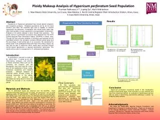
Ploidy Makeup Analysis of HypericumperforatumSeed PopulationThurman Redhouse Jr.1,3, Luping Qu2, Mark Widrlechner1. New Mexico State University, Las Cruces, New Mexico; 2. North Central Regional Plant Introduction Station, Ames, Iowa; 3. Iowa State University, Ames, Iowa Results Abstract Populations of Hypericum perforatum may include diploid, tetraploid, and hexaploid cytotypes. Tetraploid populations are by far the most common and likely the ancestral type and is capable of asexual reproduction by apomixes. A population with mixed ploidy types may affect detrimentally on seed regeneration and germplasm conservation. Analysis of the ploidy makeup is done through flow cytometry. The seeds of three H. perforatum accessions were obtained from the North Central Regional Plant Introduction Station (NCRPIS) in Ames, Iowa. Through the flow cytometry analysis of individual seed samples we will understand whether the accession contains different ploidy types and the percentage of each ploidy type in an accession. By comparing the ploidy levels of the endosperm tissue with that of the embryo tissue, we may also be able to determine which seeds were produced through normal sexual reproduction or asexual reproduction (apomixes). The results will be useful in management of H. perforatum germplasm. A) diploid (2x = 16) embryo with tetraploid (4x = 32) endosperm B) tetraploid embryo with decaploid (10x = 80) endosperm • Introduction H. perforatumis referred to mostly as St. John's wort. It works as an anti-inflammatory, antiseptic, and antiviral. In Native American tribes such as the Cherokee, the Iroquois, and the Montagnais, they have used St. John’s wort for the treatments of fever, cough, nosebleed, snakebites and intestinal problems. Although research on medicinal plants focuses mainly on chemical properties of the plants, research on the genetics and germplasm are being done for future aid towards the success of the medicinal plants. C) hexaploid (6x = 48) embryo with decaploid endosperm. Flow Cytometry All flow cytometry was performed with a BD Biosciences (San Jose, CA) FACSCanto equipped with a 488 nm laser and 610/20 emission filter. One hundred seed samples were analyzed in each accession. Reproductive pathways were determined by calculating the ratios of embryo and endosperm nuclei DNA contents Conclusion Hypericum perforatum accessions tested in this investigation contains mixed ploidy types. Flow cytometry is a very effective and less time consuming method for evaluation of ploidy types and can also be used in determining reproductive pathways in plants by analyzing seed samples. Materials and Methods The H. perforatum seeds are obtained from the NCRPIS. Each individual seed was placed in a 5 mL tubes. An amount of 1 mL of nuclei stabilizing buffer (15 mM HEPES, 1 mM EDTA, 80 mM KCl, 20 mM NaCl, 300 mM Sucrose, 0.2% Triton X-100, 0.5 mM Spermine tetrahydrochloride, 0.25 mM PVP) was applied and the seed was broken down with a homogenizer, (Omni International 1000; Waterbury, CT.) in an effort to release the nuclei within the seed. Again 1 mL was added and then filtered. After filtration the solution was then centrifuged for 6 minutes at a speed of 800O rpm. With the nuclei settled at the bottom of each tube, the supernatant was discarded and re-suspeded with 250 µL of staining buffer(10 mM MgSO4, 50 mM KCl, 5 mM HEPES, 0.1% DL-Dithiothreitol, 2.5% Titon X-100, 100 mg/mL Propidium Iodide). The solutions were then read through a flow cytometry where the stained nuclei will be measure in terms of its florescence detected within the machine. Acknowledgments We thank the USDA-ARS, National Science Foundation, and Department of Energy for funding this work. Thank you to everyone at the George Washington Carver Internship Program. Carolyn Lawrence, Candice A.C. Gardner and everyone at the North Central Regional Plant Introduction Station, Ames, Iowa.


