Nanoplasmonic Biomolecular Imaging ( 奈米電漿子生物分子影像 )
380 likes | 542 Views
Nanoplasmonic Biomolecular Imaging ( 奈米電漿子生物分子影像 ). Shean-Jen Chen ( 陳顯禎 ) 3-30 -2006. Adaptive Photonics Lab, NCKU - Nanoplasmonic Biosensing & Molecular Imaging - Adaptive Optics for Vision Science. Outlines. Surface Plasmons & Particle Plasmons

Nanoplasmonic Biomolecular Imaging ( 奈米電漿子生物分子影像 )
E N D
Presentation Transcript
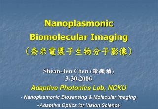
Nanoplasmonic Biomolecular Imaging(奈米電漿子生物分子影像) Shean-Jen Chen (陳顯禎) 3-30-2006 Adaptive Photonics Lab, NCKU - Nanoplasmonic Biosensing & Molecular Imaging - Adaptive Optics for Vision Science
Outlines • Surface Plasmons & Particle Plasmons • Label-free Nano-imaging: Surface Plasmon Resonance (SPR) Microscopy • Amplified Optical Near-field: Metal-tip Plasmon-enhanced Near Field Scanning Optical Microscope (NSOM) for Fluorescent or Raman Molecular Image • Breaking Diffraction Limit: Nanoplasmonic Structured Metalayer • Conclusions Adaptive Photonics Lab, NCKU
Optical Microscopy Advantages: - Intuitive image interpretation - Applicable to samples in natural environment - In general non-destructive - Easy to use Disadvantages: - Abbe diffraction limit - Large sample area exposed to illumination light
Total Internal Reflection Fluorescence Microscopy (TIRFM) • Intensity Measurement: Controlling the depth resolution via changing incident angle or wavelength Evanescent Wave Filter out scattering light “ What bother the TIRF ?” It can provide a better depth resolution than confocal microscopy, especially when it comes to selecting fluorescent molecules close to the biosensing surface. Adaptive Photonics Lab, NCKU
Outlines • Surface Plasmons & Particle Plasmons • Label-free Nano-imaging : Surface Plasmon Resonance (SPR) Microscopy • Amplified Optical Near-field : Metal-tip Plasmon-enhanced Near Field Scanning Optical Microscope (NSOM) for Fluorescent or Raman Molecular Image • Breaking the Diffraction Limit : Nanoplasmonic Structured Metamaterial • Conclusions Adaptive Photonics Lab, NCKU
Surface Plasmon Resonance (SPR) Biosensors(Kretschmann-Raether Configuration) Slide Au film Prism SAM Receptors Ligands P-wave light Detector θ Slide Au film O-ring Flow cell TE-cooler Receptor in Ligand in SAM in Ligand out SAM out Receptor out SPR condition: (Surface plasmon wave at semi-infinite structure) Adaptive Photonics Lab, NCKU
Spectrum Intensity Detection approach Angular Wavelength Intensity Phase Prism coupler 5 x 10⁻⁷ 2 x 10⁻⁵ 5 x 10⁻⁵ 2 x 10⁻⁷ Grating coupler 2 x 10⁻⁶ 6 x 10⁻⁵ 2 x 10⁻⁴ 8 x 10⁻⁷ Advantages of SPR Biosensing Theoretical sensitivity (Resolution unit: RIU; λ= 632.8 nm) • Label-free sample • Real-time biomolecular interaction analysis (BIA) • Kinetic study • Characterize and quantify biomolecular interaction • High sensitivity • (~ 1 pg/mm2) • Potential of high • throughput screening 1. ATR coupler: BK7 / Au (48 nm) / analyze (1.32). 2. Grating coupler: Pitch of 800 nm & depth of 70 nm. 3. Angular resolution is 1 x 10⁻⁴ deg. 4. Wavelength is 0.02 nm. 5. Intensity resolution is 0.2 %. 6. Phase resolution is π/200. Adaptive Photonics Lab, NCKU
Intensity and Phase Variation at SPR - Reflection relationship: - Phase variation analysis: Adaptive Photonics Lab, NCKU
Particle Plasmon Resonance (D.A. Schultz, 2003) Adaptive Photonics Lab, NCKU
Plasmonic Biosensors Four Plasmonic Effects: - Surface plasmons - Particle plasmons - Inter-particle coupling - Gap mode → To increase local EM field (about 105 times) → To increase sensitivity (less than 1 pg/mm2) 50 nm Adaptive Photonics Lab, NCKU G.-Y. Lin et al., Proc. SPIE 6095 (2006).
Au film Glass Nanoparticle-enhanced Plasmonic Biosensors • Excitation of surface plasmons and particle plasmons • Locally enhanced EM fields • Providing more sensitive biosensors • Grain size 4.0 nm & • interval 2.0 nm • Not analyte-tagged • nanoparticles • Not synthesizing W. P. Hu et al., Biosensors & Bioelectronics 19 (2004) 1465. S.-J. Chen et al., U.S. Patent Pending No. 10/660833, 2003. Adaptive Photonics Lab, NCKU
Advanced Plasmonic Biosensing What is Next? Biomolecular Imaging with Plasmonic Effects Adaptive Photonics Lab, NCKU
Outlines • Surface Plasmons & Particle Plasmons • Label-free Nano-imaging: Surface Plasmon Resonance (SPR) Microscopy • Amplified Optical Near-field : Metal-tip Plasmon-enhanced Near Field Scanning Optical Microscope (NSOM) for Fluorescent or Raman Molecular Image • Breaking the Diffraction Limit : Nanoplasmonic Structured Metamaterial • Conclusions Adaptive Photonics Lab, NCKU
Common-path Phase-Shift Interferometry SPR Imaging - Long-term stability to reject external disturbances - Easy aligned and compact system Phase-shifting Mapping to CCD Phase variation Adaptive Photonics Lab, NCKU S.-J. Chen et al., Journal of Biomedical Optics 10 (2005) 034005.
15mer DNA SPR Phase Image - Five-step Phase-shift Interferometry 0 1/2π π 3/2π 2π - Phase Reconstruction DNA DNA Adaptive Photonics Lab, NCKU Y.-T. Su et al., Optics Letters 30 (2005) 1488.
SPR Microscopy - SPR Phase Microscope for Living Cell Membrane Images with No Fluorescent Labels - Plasmon-enhanced TIR Fluorescence Microscope for Dynamic Living Cell Membrane Images ‘Surface Plasmons’ & ‘Particle Plasmons’ Adaptive Photonics Lab, NCKU
Cell Membrane Imaging Melanoma-GFP-tagged TM cell • GFP-tagged TM on the melanoma cell membrane near the chip surface is excited by the evanescent wave for TIR or surface plasmon wave for SPR; • - The enhancement of fluorescence is observed apparently btw the two images. The experimental results show that the fluorescence intensity can be enhanced about 3.0 fold. • - Because of the variant distance btw the cell membrane protein TM and the collagen-coated surface, different surface plasmon effects can be applied to interpret the phenomenon of fluorescence emission or quenching. TIRFM Plasmon-enhanced TIRFM Adaptive Photonics Lab, NCKU L.-Y. He et al., Proc. SPIE 6088 (2006). (c) Fig. 4. Cell membrane images from (a) TIRFM and (b) plasmon-enhanced TIRFM. (c) Fluorescent intensity distribution on variant pixels in white crosscut lines of above two images.
Lateral Resolution Limited by Propagation Length of Surface Plasmon Wave Propagation Length @ λ=630nm Ag = 19μm; Al = 1 μm Ag Al A Goldfish Glial Cell Interference Reflection Microscope Ag SPR Intensity Microscope Al Adaptive Photonics Lab, NCKU K.-F. Giebel et al., Biophysical Journal 76 (1999) 509.
Outlines • Surface Plasmons & Particle Plasmons • Label-free Nano-imaging : Surface Plasmon Resonance (SPR) Microscopy • Amplified Optical Near-field: Metal-tip Plasmon-enhanced Near Field Scanning Optical Microscope (NSOM) for Fluorescent or Raman Molecular Image • Breaking the Diffraction Limit : Nanoplasmonic Structured Metamaterial • Conclusions Adaptive Photonics Lab, NCKU
Near-field Scanning Optical Microscope (NSOM) • Evanescent Wave and Nano-scanning Tip Techniques to Break “Diffraction Limit” • Lateral spatial resolution: ~20nm • Longitudinal spatial resolution: ~50nm Operation: • Confined by a metal aperture • Within short distance beyond • the screen. Applications: • Single molecule to cell detection • Nanolithography • Super-resolution data storage • Near-field optical interaction on • nanoparticles, nanoclusters, • and localized surface plasmon. • Nanophotonics • Surface photochemistry Adaptive Photonics Lab, NCKU
Single-molecule Detection Liquid operation of NSOM opens the way to directly visualise and quantify the size and composition of membrane domains, like lipid rafts, in solution. Fluorescence image of a dendritic cell in buffer solution collected in confocal mode (A) and NSOM mode (B). Single molecule detection on cells by NSOM. This figure shows a 40 nm optical resolution near-field ‘zoom-in’ on the indicated area (3.2 mm2) in the bright-field image of a fibroblast expressing LFA-1-GFP. GFP excitation is accomplished using 488 nm light (Ar-Kr laser line) linearly polarized along 90°. FEBS Letters 573 (2004) 6. Journal of Cell Science 114 (2001) 4153.
Apertureless NSOM In apertureless near-field scanning optical microscopy (ANSOM), the probe vibration is often used to increase the detected signal that can be detected at by a lock-in amplifier. The realistic model of ANSOM should take into account the scan of the probe as well as the probe vibration and the material properties. Adaptive Photonics Lab, NCKU R. Fikri et al., Optics Letters 28 (2003) 2147.
Surface-Enhanced Raman Scattering (SERS) • Amplified local EM fields • To study protein conformational change • To investigate biomolecular structures C. L. Lei et al., Mater. Sci. Eng. B 32 (1995) 39. Adaptive Photonics Lab, NCKU
Metal tip-enhanced NSOM for Fluorescent Molecular Image • Enhance local EM field & improve • detecting signal • High resolution image for fluorescent • DNA samples H. Frey, PRL 93 (2004) 200801. DNA Fluorescent Image Adaptive Photonics Lab, NCKU
Metal tip-enhanced Raman Spectroscopy N. Hayazawa et al., JAP 92 (2002) 6983.
Outlines • Surface Plasmons & Particle Plasmons • Label-free Nano-imaging : Surface Plasmon Resonance (SPR) Microscopy • Amplified Optical Near-field : Metal-tip Plasmon-enhanced Near Field Scanning Optical Microscope (NSOM) for Fluorescent or Raman Molecular Image • Breaking Diffraction Limit: Nanoplasmonic Structured Metamaterial • Conclusions Adaptive Photonics Lab, NCKU
Stimulated Emission Depletion (STED) Microscopy The role of the STED beam is to induce the transition L2 L3 by stimulated emission and to deplete the excited fluorescence. Excitation pulses are followed by stimulated emission depletion pulses for fluorescence inhibition. After passing dichroic mirrors and emission filters, fluorescence is detected through a confocal pinhole by a counting photodiode. Adaptive Photonics Lab, NCKU PNAS 97 (2000) 8207.
Comparison btw Confocal & STED Reducing the fluorescence focal spot size to far below the diffraction limit: (a) spot of a confocal microscope (left) compared with that in a STED microscope (right) utilizing a y-oriented intensity valley for STED (upper right insert, not to scale) squeezing the spot in the x direction to 16 nm width. (b) The average focal spot size (squares) decreases with the STED intensity following a square-root law. Insert (right) discloses the histogram of the measured spot sizes rendering the 26 nm average FWHM. Adaptive Photonics Lab, NCKU PRL 94 (2005) 143903.
Negative Refractive Index In 1968, Veselago first considered the case of a medium that had both negative dielectric permittivity and negative magnetic permeability. Nature 420 (2002) 119. Adaptive Photonics Lab, NCKU
Negative Refraction Makes a Perfect Lens Flat Slab Object Real Image A negative refractive index medium bends light to a negative angle with the surface normal. Light formerly diverging from a point source is set in reverse and converges back to a point. Adaptive Photonics Lab, NCKU J. B. Pendry, PRL 85 (2000) 3699.
Microwave Imaging by Flat Lans using Negative Refraction Photonic Crystal at Band-gap Edge for 9.3 GHz Metal Nanostructure with square copper split ring resonators and copper wire strips The images can be observed only in a narrow frequency range, between 9.0 and 9.4 GHz. R. A. Shelby et al., Science 292 (2001) 77. P. V. Parimi et al., Nature 426 (2003) 404. Adaptive Photonics Lab, NCKU
Sub-Diffraction-Limited Optical Imaging with a Silver Superlens N. Fang et al., Science 534 (2005) 308.
Enhanced Lateral Resolution by Control of the Size & Distribution of Metal Nanoparticles • Break diffraction limit & • improve single particle • detection without near-field tip • Lateral spatial resolution: • ~100nm Enhanced the Readout Signal of Super-RENS Disc Adaptive Photonics Lab, NCKU J.-N. Yih et al., Applied Optics 44 (2005) 3001.
White beam & normal incident (TM) Λ=380nm n1 / H2O biolayer: 10nm / 1.46 0.5Λ Metal layer (δ = 60nm) grating layer (δ = 25nm) guided-layer ( d = 250 nm) n2 / Ta2O5 n3 / BK7 substrate (1000μm) Coupled Waveguide-surface Plasmon Resonance Biosensors Constructed with Sub-wavelength Grating Red line: without a bio-layer Blue line: with a bio-layer 21.2 nm • The reflectivity spectrum of CWSPR biosensors with sub-wavelength grating • Localized surface plasmon mode and waveguide mode • Sharper dip-width • Enhance sensitivity Adaptive Photonics Lab, NCKU
EM Wave transport below the Diffraction Limit in Metal Particle Plasmon Waveguides (S. A. Maier et al., 2003) Adaptive Photonics Lab, NCKU
Anticipated Research Outcomes • Establish the immobilization of biomolecules • Build the real-time portable SPR and SERS metrology systems • Using plasmonic biosensors to achieve the direct detection of drug • Conformational & Structural information from CWSPR & SERS • A total solution of the operation of advanced plasmonic biosensing • Single Bio-molecule Detection for BIA • Molecular Scale Imaging of Living Cell Adaptive Photonics Lab, NCKU