Download
1 / 38
390 likes | 578 Views
Origin of signals in tissue imaging and spectroscopy. Andrew J. Berger The Institute of Optics University of Rochester Rochester, NY 14627. A very brief outline. Absorption Emission Scattering. Who are you? Why are you here?. (with apologies to Admiral Stockdale).

E N D
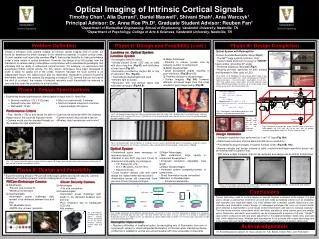
Origin of signals in tissue imaging and spectroscopy Andrew J. Berger The Institute of Optics University of Rochester Rochester, NY 14627
A very brief outline • Absorption • Emission • Scattering
Who are you? Why are you here? (with apologies to Admiral Stockdale) • experienced in some branch of optics • biomedical not your main shtick • interested in survey of fundamentals • want introduction to applications • interested in following the later talks • want pointers to the literature
Fred the photon photons absorption events Absorption = molecular transition between states • electronic • vibrational • rotational • (translational)
outer shell: n>1 13.7 eV = 91 nm Electronic transitions What's quantized: energy 4 Consequently: 3 2 1 Biologically: typically UV or blue
Representative values: mid-IR Vibrational transitions What's quantized: energy
Representative values: microwave regime Rotational transitions What's quantized: Consequently:
How to talk about absorption molar extinction "absorption coefficient" [1/length] concentration
What's absorbing? DNA biologicalwindow rotational vibrational electronic courtesy V. Venugopalan, http://www.osa.org/meetings/archives/2004/BIOMED/program/#educ
Hemoglobin courtesy V. Venugopalan, http://www.osa.org/meetings/archives/2004/BIOMED/program/#educ
blood = 45% red blood cells by volume red blood cell = 1/3 hemoglobin by weight Hemoglobin molecular weight = 65,000 mg/mmole Typical tissue absorption! adipose tissue ~ 1% blood by volume Hb concentration = 23 mM
Hemoglobin at isosbestic point, Mean free absorption pathlength = 500 mm (!)
Hemodynamics calculations single absorber : two absorbers : measure the absorption coefficients look up the molar extinction coefficients (e.g. http:/omlc.ogi.edu) calculate the concentrations oxygen saturation: parameters of interest : theory works for N>2 chromophores, too! total hemoglobin
Further adventures of Fred the photon absorption photons fluorescence
Fluorescence: level diagram • absorption: fsec • internal conversion: fsec • upper state lifetime:psec-nsec • emission: fsec shift is to the RED (Stokes) of the excitation light
Fluorescence Spectroscopy Major biological fluorophores: • structural proteins:collagen and elastin crosslinks • coenzymes for cellular energy metabolism (electron acceptors): • flavin adenine dinucleotide (FAD) • nicotinamide adenine dinucleotide, reduced form (NADH) • aromatic amino acids: side groups on proteins • porphyrins: precursors to heme courtesy M.-A. Mycek Ref. Mycek and Pogue, Handbook of Biomedical Fluorescence
A fluorescence scenario cellular epithelium thickening collagen support healthy trending towards cancer • increased FAD fluorescence • reduced collagen fluorescence (farther from surface) • polyp formation → neovasculature; increased absorption & decreased fluorescence
The time dimension • absorption: fsec • internal conversion: fsec • upper state lifetime:psec-nsec • emission: fsec • radiative decay rate:kr • nonradiative loss rate:knr • knr varies with environment • fluorescence decay lifetime varies, too: not intensity-based! combined spectral and temporal fluorescence measurements: Pitts and Mycek, Rev. Sci. Inst.72:7, 3061-3072 (2001).
More introductions to fluorescence R. Redmond, "Introduction to fluorescence and photophysics," in Handbook of Biomedical Fluorescence (ed. Mycek and Pogue). N. Ramanujam, "Fluorescence spectroscopy of neoplastic and non-neoplastic tissues,"Neoplasia, 2:1, 89-117 (2000).
Yet more adventures for Fred scattering photons Stokes Anti-Stokes Raman scattering
incident photon has energy E molecule gains energy DE scattered photon has energy E -E Level diagram for Raman energy excitation usually in near-IR or <300 nm UV to avoid visible fluorescence
Basic mechanism of Raman scattering induced dipole moment : product term : STOKES ANTI-STOKES
Typical spectrum (oral bacteria) 1005 1092 619 783 667 720 902 1457 853 1340 1127 813 1259 1211 guanine phenylalanine tyrosine adenine intensity (arb. units) C-N, C-C str. 1651 cytosine, uracil phenylalanine amide III C-H 2 def. 1580 amide I aromatic amino acids RNA bases Raman shift (cm-1)
Applications for Raman • Chemical analysis of tissue, in vitro or in vivo (breast, artery, blood) • Disease classification • High-resolution, molecularly specific microscopy topical review: Hanlon et al., “Prospects for in vivo Raman spectroscopy,” Phys. Med. Biol. 45, R1-R59 (2000) (or just talk to me!) go to: FWN4, “CARS microscopy: coming of age,”Sunney Xie, 2:45-3:15. FWN5, “Interferometric contrast between resonant CARS and nonresonant four-wave mixing,”Daniel Marks, 3:15-3:30.
Fred keeps going, and going, and... scattering photons elastic scattering
caused by variations in refractive index Elastic scattering componenttypical n in the vis/NIR extracellular fluid1.35 – 1.36 cytoplasm1.36 – 1.375 nucleus1.38 – 1.41 mitochondria1.38 – 1.41 water1.33 Drezek et al., Appl. Opt. 38:16, 3651-3661 (1999). • various approaches to modeling: full rigorMaxwell’s equations (e.g. Drezek above) Mie theoryplane wave on homogeneous sphere (e.g., code at philiplaven.com) van de Hulstthree-term approximation to Mie (larger spheres and modest n values) Rayleigh scatteringvery small particles (compared to λ)
Polystyrene Spheres of Varying Diameters in Water 0 10 ) -1 Mie Theory Scattering Coefficient (mm 2000 nm -1 1000 nm 10 200 nm 100 nm 20 nm -4 l 500 600 700 800 900 1000 1100 Wavelength (nm) Wavelength dependence varies w/ scatterer size courtesy Edward Hull, Rochester summer school lecture notes
A summary of scattering scales Figure by Steve Jacques, Oregon Medical Laser Center http://www.omlc.ogi.edu/classroom go to: FTuL1, “On the microscopic origin of light scattering in tissue,”Peter Kaplan, 2:00-2:30.
d/2 (F = cavity finesse) etalon Spectral dependence of scattering incident plane wave van de Hulst approximation to Mie theory sphere d
Spectral dependence of scattering 1-D etalon • d=5 microns • n1 = 1.36 • n2/n1 = 1.06 3-D sphere wavelength / nm
superposition of spectra mixture Scattering spectroscopy more rapid oscillations • spacing of peaks:size of scatterer • depth of modulation:number of such scatterers
Scattering spectroscopy broadband polarized illumination polarization-resolved detection normal colon cells cancerous cells Perelman et al., Phys Rev Lett80:627 (1998) and following.
Angularly-resolved scattering d angular distribution has interferometric (oscillatory) behavior as well go to: FTuR1, “Real-time angle-resolved low-coherence interferometry for detecting pre-cancerous cells,”Adam Wax, 4:15-4:45. FTuL4, “Elastic-scattering spectroscopy for cancer detection: What have we learned from preliminary clinical studies?”Irving Bigio, 3:00-3:30.
Bulk tissue interrogation reduced scattering coefficient [1/length] • determine the absorption coefficient (spectroscopy) • identify and characterize heterogeneities (functional imaging) • note: scattering enables absorption studies in backscattering geometry!
absorption RMS distance from origin (“random walk”) increases according to no absorption pulse diffusion coefficient [m2/sec] scattering Absolutely basic photon migration in the limit of: signal at detector decays according to Detector no scattering
different source-detector separations ma = 0.001 mm-1 35 mm ms' = 1 mm-1 n = 1.4 25 mm r = 15 mm The real deal: diffusion theory scattering and absorption pulse
time domain: intensity vs. time frequency domain (amplitude-modulation): modulation depth and/or phase vs. distance or frequency steady state: intensity vs. distance What are the diffusion measurements? source(s) detector(s) go to: FTuK1, “Multidimensional diffuse optical imaging in breast cancer detection,”Brian Pogue, 2:00-2:30. FTuK5, “Functional imaging by optical topography,”Randall Barbour, 3:15-3:45.
Still hungry? • fluorescence:multiphoton-excited microscopy • second-harmonic: ditto • elastic scattering:optical coherence tomography, laser scanning confocal microscopy • polarization:surface-sensitive imaging, intrinsic birefringence • instrumentation: Raman fiber probes, fluorescence excitation-emission matrices Thanks to: Mary-Ann Mycek, Vasan Venugopalan, Edward Hull Have a great rest of the conference!