Download
1 / 22
550 likes | 1.66k Views
Introduction to EPR/ESR Spectroscopy and Imaging. Suggested reading: C.P.Poole, Electron Spin Resonance, A comprehensive Treatise on Experimental Techniques. J.A.Weil, J.R.Bolton, J.E.Wertz, Electron Paramagnetic Resonance: Elementary Theory and Practical Applications.

E N D
Introduction to EPR/ESR Spectroscopy and Imaging Suggested reading: C.P.Poole, Electron Spin Resonance, A comprehensive Treatise on Experimental Techniques J.A.Weil, J.R.Bolton, J.E.Wertz, Electron Paramagnetic Resonance: Elementary Theory and Practical Applications G.R.Eaton, S.S.Eaton, K.Ohno, EPR imaging and In vivo EPR
Magnetic momentum of an add electron s = gS N L = gL N = 1838 This is the ratio of rest mass of proton to the rest mass m of electron Thus EPR energies are generally about 2000 times as big as NMR energies
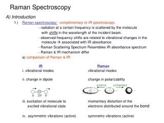
NMR – EPR comparison of energies NMR Radio wave in the range : 90 – 700 MHz Field value : 2 - 14 T Relaxation time : 10-3 to 10 sec EPR Microwave in the range : 1.2 GHz – 100 GHz Field : 0.03 – 0.3 T Relaxation time : 10-9 – 10-6 sec “Additional problems with biological EPR spectroscopy is the microwave absorption H2O in biological objects.”
Principle of EPR spectroscopy Relaxation T1 – Spin lattice relaxation E = g(B0+B1) T2 – Spin-spin relaxation T2* – Spin-spin relaxation B0 Expt. Obtained spectrum Absorption spectrum
Field (B1) modulation in EPR Why: Absorption signal is weak, compared NMR, and buried under equally amplified noise. Modulation frequency Modulation amplitude Oscillating Magnetic field B1 Unmodulated Modulated
5 5 1 1 4 4 2 2 3 3 Phase Sensitive Detection in EPR Max 0 -Max Field Field
-1 0 1 + 2 +1 -1 1 - 2 0 +1 N S = 1 for 14N O. 2S+1 = 3 Nuclear magnetic coupling – “Hyperfine splitting”
+1 0 1 + 2 -1 1 1 + + 2 2 +1 1 1 1 1 - - + + 2 2 2 2 1 1 1 1 - - + + 0 1 2 2 2 2 - 1 1 2 -1 - - 2 2 Expected Experimentally measured N O. Secondary Hyperfine Splittings H
EPR spin trapping Many free radicals, generated by enzymatic reactions are not stable enough to detect by EPR spectroscopy. • Superoxide radical (O2.-) • Hydroxyl radical (OH.) • Nitric oxide (NO:) They need to be stabilized to detect by EPR: “Spin trapping” Spin trap + Unstable radical Stable radical (?) (No EPR signal) (No EPR signal) (EPR signal)
Xanthine xo Hypoxanthine O2 O2-. DEPMPO + DEPMPO-OOH Superoxide trapping: Example 1 Xanthine / Xanthine oxidase EPR spect. of DMPO-OH
Trapping Nitric Oxide Although NO is paramagnetic, it is impossible to detect by EPR directly, because being small, it relaxes very fast as in the case of O2. Thus special approaches are required to restrict its motion to get reasonable spectrum. Fe complexes of dithiocarbamate and its derivatives
Fe(MGD) Fe(MGD)-NO
Superoxide trapping: Example 1 Nitric oxide synthase (NOS) Fe-MGD DMPO-OO-
EPR Imaging – Concept of gradient Field 1 2 MAGNET MAGNET 4 3 Bo Field is being uniform (g(B0+B1)) all the four spin pockets come to resonance frequency at a time
1- 4 Gradient generation 1 2 3 4 Bo Bo Bo Projections 1 2 2, 4 1, 3 N S 3 4 Bo (x+Bo) (x-Bo) Bo Re-construction 1,4 x-Bo 2 3 1 2 N S 3 4 x+Bo 2D image Principle of cw EPR Imaging Projection Gradient Direction N S
Pros and Cons of EPR imaging • Not adequate concentration of radicals available in biological systems • Needs exogenous infusion of stable radicals species in organs or whole body imaging • Needs significant reduction of microwave frequency to avoid microwave absorption. This significantly compromises the sensitivity But…. • It is an unique technique to study redox status of tissues, organs or in whole body, which cannot be achieved by other techniques
256 0 RESONATOR 3.0 4.5 6.0 7.5 9.0 10.5 12.0 13.5 15.0 16.5 NORMAL TISSUE RIF-1 TUMOR Time (min) Kuppusamy et al, Canc. Res, 1998, 58, 1562
Nitroxide intensity -> 3-CP room air 40 3-CP Carbogen 30 15N-TPL room air Frequency 20 10 15N-TPL Carbogen 0 0.05 0.10 0.0 0.15 Rate constant (min-1) Pharmacokinetics of Nitroxides at different Oxygenation of RIF-1 Tumor Carbogen Breathing Mouse (pO2= 95 mmHg) Room air Breathing Mouse (pO2=2.5 mmHg) 15N-TPL and LiPc 0.5 min 10 min Nitroxide intensity -> 100 60 40 Frequency 10 I/I0 x 100 20 0 0.0 0.05 0.10 0.15 1 Rate constant (min-1) 30 0 10 20 40 Time (minutes) Ilangovan, G. et al Mol. Cell. Biochem.,2002, 234, 393
Example 1 In vivo Imaging of NO generation Fe-MGD + NO Fe-MGD-NO No EPR signal No EPR signal Strong EPR signal NO generated in the thoracic region of a mouse, subjected to cardiopulmonary arrest