Motor Areas
530 likes | 779 Views
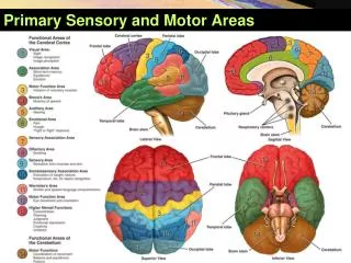
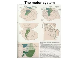
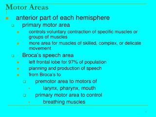
Motor Areas. anterior part of each hemisphere primary motor area controls voluntary contraction of specific muscles or groups of muscles more area for muscles of skilled, complex, or delicate movement Broca’s speech area left frontal lobe for 97% of population

Motor Areas
E N D
Presentation Transcript
Motor Areas • anterior part of each hemisphere • primary motor area • controls voluntary contraction of specific muscles or groups of muscles • more area for muscles of skilled, complex, or delicate movement • Broca’s speech area • left frontal lobe for 97% of population • planning and production of speech • from Broca’s to • premotor area to motors of • larynx, pharynx, mouth • primary motor area to control • breathing muscles
Association Areas • often adjacent to primary sensory areas • usually receive input from both primary sensory areas and other brain regions • integrate sensory experiences to generate meaningful patterns of recognition and awareness • damage to primary visual area of brain results in loss of vision • damage to visual association area would not recognize what they are seeing
Association Areas • somatosensory association area • integrates and interprets sensations • determines shape and texture of object without looking at it • determines orientation of one object with respect to another as they are felt • sense relationship of one body part to another • storage of memories of past experiences to compare current sensations with previous experiences • visual association area • receives sensory impulses from primary visual area and thalamus • relates present and past visual experiences • essential in recognizing and evaluating what is seen
Association Areas • auditory association area • allows recognition of sound as speech, music, or noise • Wernicke’s area • broad region in left temporal and parietal lobes • interprets meaning of speech by recognizing spoken words • translates words into thoughts • right hemisphere correspond to Broca’s and Wernicke’s in the left • contribute to verbal communication by adding emotional content like anger, joy, or spoken words
Association Areas • common integrative area • integrates sensory interpretations from all sensory association areas • allowing formation of thoughts based on variety of sensory inputs • transmits signals to other parts of brain to cause appropriate response • premotor area • immediately anterior to primary motor area • controls learned, skilled, motor activities of a complex and sequential nature • causes specific groups of muscles to contract in specific sequence • serves as memory bank for specific patterns of movements
Association Areas • frontal eye field area • controls voluntary scanning movements of eye • reading for example
Hemispheric Lateralization • functional asymmetry • each hemisphere also specializes in performing certain unique functions • appears at about 30 weeks in fetal development • left hemisphere receives somatic sensory signals from controls muscles on the right side of the body • right hemisphere receives and controls the left
Hemispheric Lateralization • left hemisphere • reasoning, numerical and scientific skills • spoken and written language • ability to use and understand sign language • right hemisphere • musical and artistic awareness • spatial and pattern perception • recognition of faces • emotional content of language • discrimination of different smells • generating mental images of sight, sound, touch, taste, and smell to compare relationships among them
Diencephalon • extends from brain stem to cerebrum • surrounds third ventricle • thalamus • hypothalamus • pineal gland
Thalamus • 3cm in length • 80% of diencephalon • paired oval masses of gray matter • organized into nuclei • interspersed tracts of white matter • intermediate mass • crosses third ventricle • joins left and right halves of thalamus • relays and processes sensory and motor information
Thalamus • with other parts of the brain helps regulate • autonomic activities • emotions • maintains consciousness • pain perception • learning • memory • cognition (thinking and knowing)
Hypothalamus • small part of diencephalon inferior to thalamus • 12 or so nuclei • mammillary bodies • serve as relay stations for reflexes to sense of smell • infundibulum • connects pituitary gland • major regulator of homeostasis • continually monitors conditions within blood • osmotic pressure, glucose levels, hormone concentrations, temperature
Important Functions of Hypothalamus • Control of ANS • regulator of visceral activities including heart rate, movement of food through GI tract, contraction of urinary bladder • Production of hormones • through bloodstream and axons to pituitary • Regulation of emotional and behavioral patterns • works with limbic system • Regulation of eating and drinking • insulin passes BBB stimulating or inhibiting food intake • thirst center sensitive to osmotic pressure
Important Functions of Hypothalamus • Control of body temperature • stimulates body to promote heat loss if too hot and heat production and retention if too cold • Regulation of circadian rhythms and states of consciousness • sleep and wake cycles
Pineal Gland • size of a small pea • considered part of endocrine system • secretes melatonin • promotes sleepiness • contributes to setting of body’s biological clock
Brain Stem • between spinal cord and diencephalon • three regions • midbrain • pons • medulla oblongata • contain both tracts and nuclei • act as relay centers for processing and controlling • involuntary of sight and sound processing • eye movement • regulation of autonomic functions • respiration, heart rate, blood pressure, digestion
Midbrain • conducts nerve impulses from cerebrum to spinal cord, medulla, and pons • peduncles have axons of sensory neurons • medulla to thalamus • regions • corpora quadrigemina • superior colliculi • reflex centers for some visual activities • inferior colliculi • reflex centers for some reactions to auditory stimuli
Pons • anterior to cerebellum • inferior to midbrain • 2.5cm long • tracts that connect parts of brain • e.g. left and right cerebellum • voluntary movements relayed from cerebral cortex to cerebellum • help control breathing • pneumotaxic area • apneustic area
Medulla Oblongata • inferior part of brain stem • continuation of spinal cord • 3cm from pons to foramen magnum • all sensory and motor tracts connecting brain and spinal cord • pyramids • white matter bulges • largest motor tracts • 90% of left pass to right & right to left • cardiovascular and medullary rhythmicity areas
Reticular Formation • small clusters of neuronal cell bodies within small bundles of myelinated axons • RAS • sensory axons that project cerebral cortex • helps maintain consciousness • active during awakening from sleep • RAS arouses cerebral cortex • motor tracts • help regulate muscle tone
Functions of Cerebellum • anterior and posterior lobes govern subconscious aspects of skeletal muscle movements • flocculondular lobe on inferior surface contributes to equilibrium and balance • main function • cerebellum evaluates movements • smoothes movements • corrects errors • coordinates sequence • regulates posture and balance • makes possible all skilled muscular activities
External Spinal Cord Anatomy • roughly circular but flattened slightly in anterior-posterior dimension • ~2cm diameter • larger in cervical and lumbar enlargements • smallest at inferior tip • adult spinal cord length • from 42 to 45 cm • from medulla oblongata to L2 • filum terminale • extension of pia mater • extends inferiorly • anchors spinal cord to coccyx
External Spinal Cord Anatomy 31 pair of spinal nerves • exit vertebral column at intervertebral foramina • each pair called spinal segment • paths of communication • roots • bundles of axons that connect peripheral nerves and spinal cord • posterior or dorsal root • sensory neuron axons • anterior or ventral root • motor neuron axons
Internal Spinal Cord Anatomy right and left sides • anterior median fissure • deep and wide groove • posterior median sulcus • shallower, narrower • gray matter surrounded by white matter • gray matter shaped like an “H” or a butterfly • gray commissure • crossbar of H connects gray matter L & R sides • central canal • contiguous with 4th ventricle • white commissure • anterior to gray • commissure connects white matter on L & R sides
Internal Spinal Cord Anatomy • gray matter • cell bodies, neuroglia, unmyelinated axons of sensor neurons, dendrites of interneurons, motor neurons • divided into horns • anterior horn • cell bodies of somatic motor neurons and motor nuclei • posterior horn • somatic and autonomic sensory nuclei • lateral gray horns • absent in cervical segments • cell bodies of autonomic motor neurons regulating smooth and cardiac muscle, and glands
Internal Spinal Cord Anatomy • white matter • bundles of myelinated and unmyelinated axons of sensory neurons, interneurons, and motor neurons • divided into columns • anterior, posterior, and lateral • tracts having common origin or destination and carrying similar information over long distances • sensory tracts • toward CNS • motor tracts • away from CNS
Sensory Tracts • spinothalamic tracts and posterior columns • lateral and anterior spinothalamic tracts • pain, warmth, coolness, itching, tickling, deep pressure, crude poorly localized sense of touch • right and left posterior columns • proprioception, discriminative touch, two point discrimination, light pressure and vibration sensations