Download
1 / 48
840 likes | 2k Views
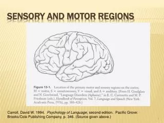
Primary Sensory and Motor Areas. Peripheral Nervous system. Peripheral nerves Cranial nerves Spinal nerves Receptors Ganglia. Peripheral Nervous system. Ganglia Autonomic ganglia Sympathetic and parasympathetic ganglia Sensory ganglia

E N D
Peripheral Nervous system • Peripheral nerves • Cranial nerves • Spinal nerves • Receptors • Ganglia
Peripheral Nervous system • Ganglia • Autonomic ganglia • Sympathetic and parasympathetic ganglia • Sensory ganglia • All nerves having sensory fibers also have sensory ganglia. • Therefore, all spinal nerves have sensory ganglia (31 pairs of ganglia called the dorsal root ganglion or spinal ganglion). • Cranial nerves having sensory fibers also have sensory gangliai.e. trigeminal nerve has trigeminal ganglion.
Nervous System Nervoussystem is dividedintotwofunctionalparts • Somaticnervoussystem • Relatedwiththesensationscomingfromexternalenvironmentandlocomotorsystem, andcontrollingtheskeletalmuscles. • It is consiciousandvoluntary. • Autonomicnervoussystem • Relatedwiththesensationscomingfromtheinternalenvironment (organsandsystems in the body), andcontrollingthefunction of organsandsystems • It is unconscious and involuntary.
Receptors • The sensations from the outside and inside environment are received by special sensory nerve endings or receptors. • Receptors can be classified in 5 basic types: • Mechanoreceptors • Thermoreceptors • Nociceptors • Photoreceptors • Chemoreceptors and baroreceptors
The sensory System • Sensory Receptors: • Sensory receptors receive input, generate receptor potentials, and with enough summation, generate action potentials in the neurons they are part of or synapse with. • There are 5 types based on the type of stimuli they detect: • Mechanoreceptors • Thermoreceptors • Pain receptors • Chemoreceptors • Photoreceptors
The sensory System • There are 5 types based on the type of stimuli they detect: • Mechanoreceptors - pressure receptors, stretch receptors, and specialized mechanoreceptors involved in movement and balance. • Thermoreceptors - skin and viscera, respond to both external and internal temperature • Pain receptors - stimulated by lack of O2, chemicals released from damaged cells and inflammatory cells • Chemoreceptors - detect changes in levels of O2, CO2, and H+ ions (pH) as well as chemicals that stimulate taste and smell receptors • Photoreceptors - stimulated by light
The sensory System • General senses: • A. Proprioceptors • Stretch receptors located in joints, ligaments, and tendons (respond to either stretch or compression) • Muscle spindles – modified muscle fibers with sensory nerve endings. Detect stretch and stimulate a reflex contraction • Purpose – maintain some degree of continuous contraction (partial sustained contraction) or muscle tone
The sensory System • General senses: • B.Cutaneous Receptors • Touch receptors:fine touch • Meissner’s corpuscle – fine touch, discrimination; found concentrated in places where you need to have a lot of responsiveness to a little input. • Merkel disks - found deep at the junction of the epidermis and dermis. • Root hair plexus - at the base of hair follicles.
The sensory System • General senses: • B.Cutaneous Receptors • Touch Receptors: pressure sensitive • Ruffini’s endings and Krause's end bulbs – encapsulated pressure sensors, dermis (and elsewhere), respond to continuous pressure • Pacinian corpuscles – deep pressure sensors, onion shaped capsule (layers of Schwann cells enclosed in a connective tissue membrane), respond to on-off pressure or vibration • Temperature • Free nerve endings, some responsive to heat and others responsive to cold • Pain • Free nerve endings, respond to chemicals released from damaged tissues.
The sensory System • General senses: • C.Pain Receptors • Somatic nociceptors • From skin and skeletal muscle • Visceral nociceptors • Receptors that help maintain internal homeostasis • Respond to stretch, lack of O2, chemicals released from damaged cells and inflammatory cells. • Referred pain – visceral pain afferents travel along the same pathways as somatic pain afferents, so sometimes the brain interprets the visceral pain as the more common somatic pain. Example – Often pain from the heart felt during a heart attack is perceived as a pain that originates in the left arm.
The sensory System • Special senses:They sense external stimuli. • Vision • Hearing • Taste • Smell • Equilibrium
The sensory System • Special senses:They sense external stimuli. • Chemical senses • Taste (gustation) • Smell (olfaction) • Vision • The ear • Hearing • Equilibrium
Types of Neuron Fibers • General somatic Afferent (GSA) • Sensations arising from skin receptors for touch, pain, temperature • Sensations arising from muscle spindles, golgi tendon organs, joint receptors • General visceral Afferent (GVA) • Sensations arising from organs • General somatic Efferent (GSE) • To skeletal muscles • General visceral Efferent (GVE) • To organs • Special somatic Afferent (SSA) • Vision, audition, equilibrium • Special visceral Afferent (SVA) • Smell and taste • Special visceral Efferent (SVE) • To muscles having the developmental origin from 3rd and 4th branchiomeric arch • e.g. muscles of pharynx and larynx, muscles of facial expression, muscles of mastication, muscles in the middle ear
Synapsis • The point at which the nerve impulse passes from one neuron to the another
Sensation • Conscious and subconscious awareness of changes in the external or internal environment. • Components of sensation: Stimulation of the sensory receptor → transduction of the stimulus → generation of nerve impulses → integration of sensory input.
Classification of Sensory Receptors • General senses: somatic and visceral. Somatic:Tactile, thermal, pain and proprioceptive sensations. Visceral:Provide information about conditions within internal organs. • Special senses:Smell, taste, vision, hearing and equilibrium or balance.
Types of Sensory Receptors • Free nerve endings: pain and thermoreceptors. • Encapsulated nerve endings: pacinian corpuscles. • Separate cells: hair cells, photoreceptors and gustatory receptor cells.
Generator Potential and Receptor Potential • Generator potential is produced by free nerve endings, encapsulated nerve endings, and olfactory receptors. When it reaches a threshold, it triggers one or more nerve impulses in the axon of a first-order sensory neuron. • Receptor potential triggers the release of neurotransmitter → postsynaptic potential → action potential.
Sensory Receptors and their Relation-ship to First-Order Sensory Neurons
Classification of Sensory Receptors Based on the Location • Exteroceptors • Interoceptors • Proprioceptors
Classification of Sensory Receptors based on the type of Stimulus • Mechanoreceptors • Thermoreceptors • Nociceptors • Photoreceptors • Chemoreceptors • Osmoreceptors
Adaptation of Sensory Receptors • Rapidly adapting receptors: receptors that detect pressure, touch and smell. • Slowly adapting receptors: receptors that detect pain, body position, and chemical composition of the blood.
Somatic Sensations • Sensory receptors in the skin (cutaneous sensations), muscles, tendons and joints and in the inner ear. • Uneven distribution of receptors. • Four modalities: tactile, thermal, pain and proprioceptive.
Tactile Sensations • Include touch, pressure, vibration, itch and tickle. • Tactile receptors in the skin are Meissner corpuscles, hair root plexuses, Merkel discs, Ruffini corpuscles, pacinian corpuscles, and free nerve endings.
Meissner Corpuscles or Corpuscles of Touch • Egg-shaped mass of dendrites enclosed by a capsule of connective tissue. • Rapidly adapting receptors. • Found in the dermal papillae of hairless skin such as in the fingertips, hands, eyelids, tip of the tongue, lips, nipples, soles, clitoris, and tip of the penis.
Hair Root Plexuses • Rapidly adapting touch receptors found in the hairy skin. • Free nerve endings wrapped around hair follicles. • Detect movements on the skin surface that disturb hairs.
Merkel Discs or Tactile Discs • Also known as type I cutaneous mechanoreceptors. • Slowly adapting touch receptors. • Saucer-shaped, flattened free nerve endings. • Found in the fingertips, hands, lips, and external genitalia.
Ruffini Corpuscles • Also called as type II cutaneous mechanoreceptors. • Elongated, encapsulated receptors. • Located deep in the dermis and in ligaments and tendons. • Found in the hands, and soles.
Pacinian or Lamellated Corpuscles • Large oval structure composed of a multilayered connective tissue capsule that encloses a dendrite. • Fast adapting receptors. • Found around joints, tendons, and muscles; in the periosteum, mammary glands, external genitalia, pancreas and urinary bladder.
Thermal Sensations • Thermoreceptors are free nerve endings. • Two distinct thermal sensations: • Cold receptors • Warm receptors
Pain Sensations • Protective. • Sensory receptors are nociceptors. • Free nerve endings. • Two types of pain: fast and slow. • Fast pain: acute, sharp or pricking pain. • Slow pain: chronic, burning, aching or throbbing pain.
Referred Pain • Pain is felt in or just deep to the skin that overlies the stimulated organ or in a surface area far from the stimulated organ.
Proprioceptive Sensations • Receptors are called proprioceptors. • Slow adaptation. • Weight discrimination. • Three types: muscle spindles, tendon organs and joint kinesthetic receptors.
Muscle Spindles • Interspersed among most skeletal muscle fibers and aligned parallel to them. • Measure muscle stretching. • Consists of intrafusal muscle fibers- specialized muscle fibers with sensory nerve endings and motor neurons called gamma motor neurons. • Extrafusal muscle fibers- surrounding muscle fibers supplied by alpha motor neurons.
Tendon Organs • Located at the junction of a tendon and a muscle. • Protect tendons and their associated muscles from damage due to excessive tension. • Consists of a thin capsule of connective tissue that encloses a few tendon fascicles.
Joint Kinesthetic Receptors • Found within or around the articular capsules of synovial joints. • Free nerve endings and Ruffini corpuscles in the capsules of joints respond to pressure. • Pacinian corpuscles respond to acceleration and deceleration of joints during movement.
Somatic Sensory Pathways • First-order neuron (somatic receptor to the brain stem/spinal cord) • Second order neuron (brain stem/spinal cord too the thalamus; decussate) • Third-order neuron (thalamus to the primary somatosensory area of the cortex).
Major Somatic Sensory Pathways • The posterior column-medial lemniscus pathway. • The anterolateral (spinothalamic) pathway. • The trigeminothalamic pathway. • The anterior and posterior spinocerebellar pathway.
The Posterior Column-Medial Lemniscus Pathway • Conveys nerve impulses for touch, pressure, vibration and conscious proprioception from the limbs, trunk, neck, and posterior head to the cerebral cortex.
The Anterolateral (spinothalamic) pathway • Conveys nerve impulses for pain, cold, warmth, itch, and tickle from the limbs, trunk, neck, and posterior head to the cerebral cortex.
Trigeminothalamic Pathway • Conveys nerve impulses for most somatic sensations from the face, nasal cavity, oral cavity and teeth to the cerebral cortex.
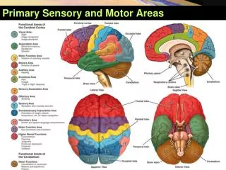
Mapping of the Primary Somatosensory Area • Mapping of the postcentral gyrus. • Size of the cortical region representing a body part depends on the sensory impulses received from that part.