Microbiology Tools
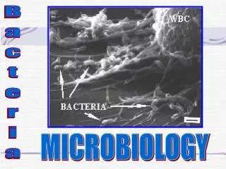
Microbiology Tools. Figure 4.1. Tools to Study the Bacteria. Different tools are employed to study bacteria Morphology Microscopy Staining . Bacterial Morphology. Three general shapes Coccus - roughly spherical Bacillus - rod-shaped Coccobacillus - short and plump


Microbiology Tools
E N D
Presentation Transcript
Tools to Study the Bacteria Different tools are employed to study bacteria Morphology Microscopy Staining
Bacterial Morphology • Three general shapes • Coccus- roughly spherical • Bacillus- rod-shaped • Coccobacillus- short and plump • Vibrio- gently curved • Spirillum- curviform or spiral-shaped • Pleomorphism- when cells of a single species vary to some extent in shape and size
Arrangement, or Grouping • Cocci- greatest variety in arrangement • Single • Pairs (diplococci) • Tetrads • Irregular clusters (staphylococci and micrococci) • Chains (streptococci) • Cubical packet (sarcina)
Arrangement, or Grouping • Bacilli- less varied • Single • Pairs (diplobacilli) • Chain (streptobacilli) • Row of cells oriented side by side (palisades)
Types of Microscopes Simple Compound Electron • Scanning Electron Microscope (SEM) • Transmission Electron Microscope (TEM)
Compound Microscope [INSERT FIGURE 4.4]
Electron Microscopy • Originally developed for studying nonbiological materials • Biologists began using it in the early 1930s • Forms an image with a beam of electrons • Electrons travel in wavelike patterns 1,000 times shorter than visible light waves • This increases the resolving power tremendously
Electron Microscopy • Magnification can be extremely high (between 5,000X and 1,000,000X for biological specimens) • Allows scientists to view the finest structure of cells • Two types: • transmission electron microscope (TEM) • scanning electron microscope (SEM)
SEM Creates an extremely detailed three-dimensional view of all kinds of objects Electrons bombard the surface of a whole metal-coated specimen Electrons deflected from the surface are picked up by a sophisticated detector The electron pattern is displayed as an image on a television screen Contours of specimens resolved with SEM are very revealing and surprising
Microscopy [INSERT FIGURE 4.13]
TEM Often used to view structures of cells and viruses Electrons are transmitted through the specimen The specimen must be very thin (20-100 nm thick) and stained to increase image contrast Dark areas of a TEM image represent thicker or denser parts
Microscopy [INSERT FIGURE 4.11]
Fixed, Stained Smears • Smear technique developed by Robert Koch • Spread a thin film made from a liquid suspension of cells and air-drying it • Heat the dried smear by a process called heat fixation • Some cells are fixed using chemicals • Staining creates contrast and allows features of the cells to stand out • Applies colored chemicals to specimens • Dyes become affixed to the cells through a chemical reaction • Dyes are classified as basic (cationic) dyes, or acidic (anionic) dyes.
Simple and Negative Staining • Simple staining: the dye sticks to the specimen to give it color • Negative staining: The dye does not stick to the specimen, instead settles around its boundaries, creating a silhouette. • Nigrosin and India ink commonly used • Heat fixation not required, so there is less shrinkage or distortion of cells • Also used to accentuate the capsule surrounding certain bacteria and yeasts
Simple Stains • Require only a single dye • A basic dye is used • Examples include methylene blue, crystal violet, basic fuchsin, and safranin • All cells appear the same color but can reveal shape, size, and arrangement
Negative Stains • Require only a single dye • An acidic dye is used • Examples include nigrosin, congo red, india ink • All cells appear clear with the background stained which reveals the shape, size, and arrangement
Differential Stains • Use two differently colored dyes, the primary dye and the counterstain • Distinguishes between cell types or parts • Examples include Gram, acid-fast, and endospore stains
Gram Staining The most universal diagnostic staining technique for bacteria Differentiation of microbes as gram positive(purple) or gram negative (red)
Gram Stain [INSERT FIGURE 4.18]
Acid-Fast Staining Important diagnostic stain Differentiates acid-fast bacteria (pink) from non-acid-fast bacteria (blue) Important in medical microbiology
Endospore Stain Dye is forced by heat into resistant bodies called spores or endospores Distinguishes between the stores and the cells they come from (the vegetative cells) Significant in medical microbiology
Special Stains Used to emphasize certain cell parts that aren’t revealed by conventional staining methods Examples: capsule staining, flagellar staining
Classification & Identification of Microorganisms [INSERT FIGURE 4.27]