SPINAL CORD ANATOMY
200 likes | 918 Views
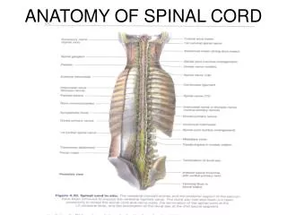
SPINAL CORD ANATOMY. General Characteristics. Approx. ½ meter in length. Varies from 1 to 1.5 cm in diameter. Enlargements: Cervical: C3 – T1. Lumbar region: L1 – S2. General Characteristics. Conus medullaris: Ends at about the level of L2. Cauda equina. Filum terminale:

SPINAL CORD ANATOMY
E N D
Presentation Transcript
General Characteristics • Approx. ½ meter in length. • Varies from 1 to 1.5 cm in diameter. • Enlargements: Cervical: C3 – T1. Lumbar region: L1 – S2.
General Characteristics • Conus medullaris: Ends at about the level of L2. • Cauda equina. • Filum terminale: Thin filament of meningeal tissue extending from conus medullaris to the coccyx.
Spinal Nerves • 08 cervical. • 12 thoracic. • 05 lumbar. • 05 sacral. • 01 coccygeal.
Plexuses • Cervical: C1 – C4. • Brachial: C5 – T1. • Lumbosacral: L1 – S4.
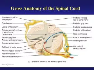
Cross-Sectional Anatomy • Central canal. • Gray matter: “H”-shaped. • White matter.
Gray Matter • Horns are equivalent to CNS nuclei. • Posterior (dorsal) horns: Receive incoming sensory fibers. • Anterior (ventral) horns: Site of cell bodies of alpha motor neurons to skeletal muscle fibers. • Lateral horns: Located only in thoracic and upper lumbar region: Site of cell bodies of ANS motor neurons.
White Matter • Columns are referred to as funiculi. • Columns consist of myelinated tracts. • Posterior (dorsal) columns. • Anterior (ventral) columns. • Lateral columns.
Commissures • White: Anterior and posterior. • Gray: Anterior and posterior.
Spinal Nerves • Dorsal rootlets. • Dorsal root: Carries afferent fibers. With dorsal root ganglion: Location of cell bodies of afferent neurons. • Ventral root: Carries efferent fibers. No associated ganglion.
Spinal Nerves • Spinal nerve carries a mixture of fibers: Both afferent and efferent. Both visceral and somatic. Proprioceptive.
Primary Rami • Dorsal (posterior): To expaxial musculature (deep back muscles) and skin on either side of dorsum of back. • Ventral (anterior): To hypaxial musculature (rest of skeletal musculature including muscles of limbs) and rest of skin.
Autonomic Rami • White ramus communicans: Carries pre-ganglionic sympathetic fibers from lateral horns of central gray of spinal cord (thoracolumbar regions). • Gray ramus communicans: Carries post-ganglionic sympathetic fibers from paravertebral ganglia back to spinal nerve.
Meniniges • Epidural space. • Dura mater: Tough outer layer. • Subdural space. • Arachnoid.
Meniniges • Subarachnoid space: Contains CSF. • Pia mater: Delicate innermost layer. • Denticulate ligaments.