SPINAL CORD ANATOMY & PHYSIOLOGY
600 likes | 1.46k Views
SPINAL CORD ANATOMY & PHYSIOLOGY. HONORS ANATOMY & PHYSIOLOGY. Spinal Cord. w/spinal nerves contain neural circuits that mediate some of your most rapid reactions to environmental changes. Protective Structures. 2 types of CT coverings surround & protect delicate nervous tissue

SPINAL CORD ANATOMY & PHYSIOLOGY
E N D
Presentation Transcript
SPINAL CORD ANATOMY & PHYSIOLOGY HONORS ANATOMY & PHYSIOLOGY
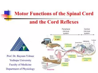
Spinal Cord • w/spinal nerves contain neural circuits that mediate some of your most rapid reactions to environmental changes
Protective Structures • 2 types of CT coverings surround & protect delicate nervous tissue • bony vertebrae • tough CT meninges, w/cushion of CSF
Meninges • 3 CT coverings that encircle spinal cord & brain: • Spinal meninges covers spinal cord • Cranial meninges covers brain
Meninges Layers: Dura Mater • “tough mother” • most superficial layer • made of dense, irregular CT • continuous with cranial meninges • forms sac from foramen magnum S2 • layer of adipose tissue between dura mater & wall of vertebral cavity (epidural space)
Middle Meninges: Arachnoid Mater • “spider-like” • deep to dura mater, superficial to pia mater • contiguous with cranial arachnoid • between dura & arachnoid = subdural space
Innermost Meninges:Pia mater • “delicate” • thin, transparent CT • adheres to spinal cord & brain • between arachnoid & pia = subarachnoid space
Spinal Tap • aka lumbar puncture • long needle inserted into subarachnoid space • adults: between L3 –L4 or L4 – L5 (inferior to lowest portion of spinal cord) • purpose: withdraw CSF for • diagnostic purposes • insert antibiotics/contrast media for myelography/ anesthetics/ chemotherapy
Spinal Cord • cylindrical with flattening of its AP diameter • adults:extends from medulla oblongata L2 vertebra • newborns: extends to L3 or L4 • elongation of spinal cord stops ~age 3-4 but growth of vertebral column continues
Spinal Cord: External View • 2 obvious enlargements noted: • cervical enlargement • C4 – T1 • serve upper limbs • lumbar enlargement • T9- T12 • serve lower limbs
Spinal Cord: External View • conusmedullaris: tapered conical structure of spinal cord below lumbar enlargement ending @ L1 – L2 • filumterminale: extension of pia mater extends inferiorly & anchors cord to coccyx • caudaequinae: “horse tail” nerves that arise from lumbar, sacral, & coccygeal portions of spine
Spinal Nerves • 31 pairs spinal nerves emerge thru intervertebral foramen • 8 pair cervical nerves: C1 – C8 • 12 pair thoracic nerves: T1 - T12 • 5 pair lumbar nerves: L1 – L5 • 5 pair sacral nerves: S1 – S5 • 1 pair coccygeal nerves: Co1
Spinal Nerves • 2 bundles of axons, called roots, connect each spinal nerve to segment of spinal cord
Spinal Cord Roots • posterior (dorsal) root • only sensory axons • each has dorsal root ganglion containing cell bodies of sensory neurons • anterior (ventral) root • only motor axons
Internal Anatomy of Spinal Cord • 2 grooves penetrate white matter & divide it in right & left sides: • anterior median fissure • deeper, wider of the 2 • posterior median sulcus • shallower, narrow furrow
Internal Anatomy ofSpinal Cord • gray matter shaped like “H” or a butterfly & is surrounded by white matter • gray commissure forms the “H” crossbar • central canal small hole in its center • extends entire length of spinal cord • filled with CSF • @ superior end is contiguous with 4th ventricle of brain
Spinal Nerves • & the nerves that branch off them are part of PNS • emerge from vertebral column thru intervertebral foramina
Spinal Nerves • typically has 2 connections to spinal cord • dorsal root (sensory) • ventral root (motor) • classified as “mixed”
Spinal Nerve Plexuses • a network of nerves (or veins, or lymphatic vessels)
Cervical Plexus • supplies skin & muscles of the head, neck, & superior portion of shoulders, chest, & diaphragm • C1 – C 5
Brachial Plexus • supplies the shoulders & upper limbs
Lumbar Plexus • supplies anterolateralabd wall, external genitals, part of lower limb
Sacral Plexus • supplies buttocks, perineum, & lower limbs
Dermatomes • cutaneous area developed from one embryonic spinal cord segment & receiving most of its sensory innervation from one spinal nerve • knowing which spinal cord segments supply each dermatome makes it possible to locate damaged regions of the spinal cord
Reflexes & Reflex Arches • reflex: a fast, automatic, unplanned sequence of actions that occurs in response to a particular stimulus • can be: • inborn • pulling hand away from hot stove • learned or acquired • foot on brake when see dog run in front of car
Pupillary Light Reflex • pupils of both eyes decrease in diameter when either eye is exposed to light • absence of a normal pupillary light refex indicates possibility of brain damage or injury
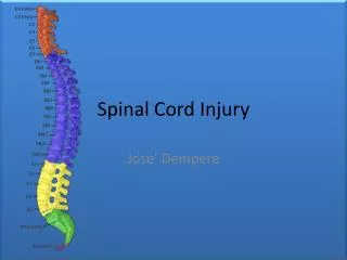
Spinal Cord Injuries • most due to trauma • cervical, lower thoracic, upper lumbar most common regions involved • paralysis • depends on location, extent of damage • monoplegia: 1 limb • paraplegia: both lower limbs • hemiplegia: upper limb, trunk, lower limb on 1 side of body • quadriplegia: all 4 limbs & trunk
Extent Muscle Paralysis • C1 – C3: no function neck down, requires ventilator to breathe • C4 – C5: diaphragm, allows breathing • C6 – C7: some arm, chest, allows breathing, moving wheelchair • T1 – T3: intact arm function • T4 – T9: control of trunk above umbilicus • T10 – L1: most thigh muscles, walk w/long leg braces • L1 – L2: most leg muscles, walk w/short leg braces
Shingles • acute infection of PNS • caused by herpes zoster (chicken pox) • virus stays in posterior root ganglion • becomes reactivated normally immune system will prevent it from spreading • reactivated virus can overcome weakened immune system leaves ganglion travels down sensory neurons supplying skin
Medical Terminology • meningitis: inflammation of meminges due to infection, bacterial (worse) or viral, vaccine protests against some bacterial causes: headache, N/V, fever, stiff neck • neuralgia: pain along a sensory nerve, trigeminal neuralgia • neuritis: inflammation of 1 or several nerves • paresthesis: abnormal sensation