Download
1 / 26
260 likes | 424 Views
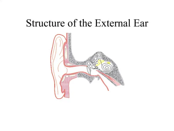
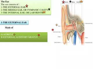
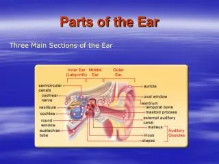
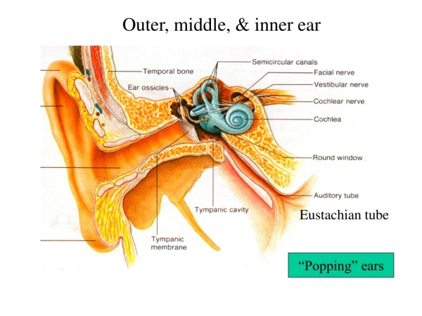
Structure of the Ear. Goldstein, pp. 343 – 360 CWE, pp. 187 – 204 Levine, pp. 339 – 346, 350 – 357, 363 – 366 Purves et al., Chapter 12. Middle ear. Outer ear. Inner ear. Auditory channel, 3cm. Functions of the external or outer ear.

E N D
Structure of the Ear • Goldstein, pp. 343 – 360 • CWE, pp. 187 – 204 • Levine, pp. 339 – 346, 350 – 357, 363 – 366 • Purves et al., Chapter 12
Middle ear Outer ear Inner ear Auditory channel, 3cm
Functions of the external or outer ear • Gathering sound energy and focusing it on the eardrum (tympanic membrane) • Selectively boosting sound pressure 30- to 100-fold for frequencies around 3000 Hertz • Pinna and concha filter different sound frequencies in order to provide cues about the elevation of the sound source (localization)
Middle ear Ossicles amplify the vibrations in two ways: • Concentrating the vibration of the large tympanic membrane onto the much smaller stapes • Create a lever action 55 mm2 3.2 mm2 ! pressure can be increased by factor 17
Lever principle Length of incus Length of malleus Fresultant = Fapplied(D1/D2) ! the force of the incoming auditory signal is increased by the ossicles by a factor of about 1.3
Reduction in sound level • Two sets of muscles in the middle ear contract and reduce the magnitude of vibration transmitted through the middle ear. • Tensor tympani! attached to the malleus ! pulls tympanic membrane ! increase in stiffness ! reducing the magnitude of vibration from incoming sounds • Stapedius muscles! connect to stapes ! retract from its normal postion ! reducing the amount of movement of the stapes • Muscles contract reflexively in response to very loud noises and can cause reductions in sensitivity by as much as 30 dB.
Inner ear 35 mm long, 2mm Ø
Scala vestibuli and scala timpaniare filled with incompressible fluid, the perilymph. • Both channels behave as one hydrodynamic system, because they are connected at the far end, or apex, by a small hole in the partition called helicotrema. • The lower section is sealed off with an elastic membrane at the round window.
The partition separating both scalae , called scala media, is filled with another fluid, the endolymph. • Its boundaries are the basilar membrane which holds the sensory organ proper (organ of corti), the Reissner's membrane, which serves to separate endolymph from perilymph, and the rigid lateral wall of the cochela.
Inner and outer hair cells about 40 cilia about 140 cilia
All of the hairs in a bundle tend to move, or bend, as a unit. • Movement of the bundle of cilia toward the tallest one increases the firing rate of the cochlear nerve axon attached to the hair cell, while movement away from the tallest one decreases it.
About 30 000 nerve fibers, whose cell bodies are located in the spiral ganglion, form connections with the bases of hair cells of each cochlea. • About 95% of them make connections with the inner hair cells at the same place where the fibers enter the cochlea. • There about 15 fibers per inner hair cell in the middle of the cochlea and about 3 to 4 per inner hair cell at the base and apex. • The remaining 5 % of the fibers from the spiral ganglion each make synaptic contact with about 10 outer hair cells located closer to the base than the fibers' point of entry into the cochlea.
Frequency Analysis in the Cochlea and Auditory Nerve • Goldstein, pp. 351 – 360 • CWE, pp. 195 – 204 (parts) • Levine, pp. 350 – 360 (parts)
Georg von Békésy 1961 Nobel Laureate in Medicine for his discoveries of the physical mechanism of stimulation within the cochlea. Born in 1899, Budapest, HungaryDied in 1972, Honolulu, Hawaii
Envelope Base Apex Displacement of the cochlear membrane in response to a 200 Hz tone. The solid curves represent the patterns of displacement at four instances; the darker the line the later in time the configuration occurred. The dashed curve is the envelope of maximum displacement.
Envelopes of vibration patterns on the basilar membrane for pure tones of different frequencies.