Download
1 / 36
410 likes | 960 Views
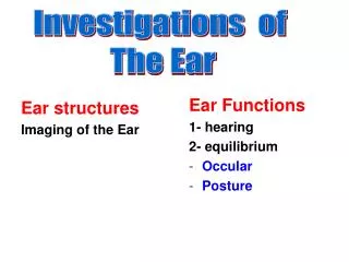
Ear structures Imaging of the Ear. Ear Functions 1- hearing 2- equilibrium Occular Posture. Investigations of The Ear. Radiology of The Ear. MRI. Plain X Ray. CT Scan. Demonstrates VIII nerve Brain Great vessels. Of limited value Demonstrates Mastoid air cells.

E N D
Ear structures Imaging of the Ear Ear Functions 1- hearing 2- equilibrium Occular Posture Investigations of The Ear
Radiology of The Ear MRI Plain X Ray CT Scan Demonstrates VIII nerve Brain Great vessels Oflimited value Demonstrates Mastoid air cells • Accurately demonstrates • External ear • Middle ear • Surrounding structures
lateralOblique (Mastoid)view Locate the • Temporo-mandibular joint • The external auditory canal (EAC) which is a complete circle • The mastoid air cells are behind and above the EAC Pneumatised mastoid: air spaces separated by bony partitions
The mastoid cells (white arrow) are obscured, and not air-containing, due to chronic otitis media. External auditory canal TMJ
Schüller view: Well-developed normally pneumatized mastoid air cells can be observed in the picture on the left side (double arrow). • In the picture on the right side, the mastoid cells (arrow) are obscured, and not air-containing, due to chronic otitis media. TMJ Sinodural angle EAC
External auditory canal TMJ • There is a clean cavity behind and above the external auditiry canal not surrounded by sclerosis • Diagnosis: surgical cavity of mastoidectomy
Axial CT scan, the destructed apex of the petrous bone can be observed (white arrow), which is caused by ? cholesteatoma. Petrous bone
Axia CT scans: • The mastoid cells on the right side (green arrow) are totally obscured, which proves mastoiditis. • On the left side (blue arrow), an intact status can be seen.
Axial CT scans: Transverse temporal bone fracture (arrows).
Investigations of Ear Functions 1- Hearing 2- Equilibrium - Occular - Posture
Investigations of Hearing Each sound has ; • Intensity : شدة الصوت • Frequency تردد الصوت • لاحظ أن: • صوت الرجل غليظ لأن تردده منخفض بينما يكون الصوت حادا في الاطفال والاناث حيث يكون تردد الصوت مرتفع
. Objective لا يتطلب تعاون بين المريض والطبيب يتم قياس الاستجابة اللاارادية للمؤثرات الصوتية We have 2 types of audiometric tests Subjective لابد من تعاون المريض مع الطبيب يعتمد الطبيب علي اجابة المريض اذا كان يسمع الصوت ام لايسمعه Pure tone audiometry Speech audiometry -Tympanometry -Electric Rersponse audiometry -Otoacoustic emmision
Pure Tone Audiometry The examiner measures The Thresholdof hearing for different frequencies, 250 Hz, 500, 1000, 2000, 4000 and 8000 Hz The sound is delivered through - Head Phone For AIR CONDUCTION and, - Bone Vibrator for BONE CONDUCTION The results are recorded on a graph (pure tone audiogram)
Value : Useful for diagnosis of: 1- Degreeof HearingLoss ( mild, moderate severe or profound) 2-Type of Hearing loss, ( CD, SNHL Or Mixed) How?
Degree of Hearing Loss Hearing loss : Up to 20 dB 20 to 45 dB 45 to 55 dB 55 to 75 dB 75 to 100 dB Above 100 dB
Type of Hearing Loss • This audiogram shows normal hearing. Air conduction & bone conduction are at normal range i.e below 20 dB
This audiogram shows a typical picture of conductive deafness in a child’s left ear. Bone Conduction Normal at the range below 20 dB Air Conduction ِ A problem in sound conduction Notice the Air Bone Gap So this is an audiogram of conductive deafness
Mixed conductive and sensorineural hearing loss Bone Conduction A problem in sound perception Air Conduction A problem in sound conduction Notice The Air Bone Gap So, this is an audiogram of Mixed Hearing Loss
The audiogram here shows a severe SN hearing loss. A problem in sound perception for Air conduction & Bone conduction NO AIIR BONE GAP
Measures the patient ability to hear & understand words لسته من الكلمات يتلقاها المريض من الطبيب بواسطة ميكرفون الي سماعة الرأس التي يضعها المريض علي رأسه ويطلب الطبيب من المريض أن يكرر الكلمات Speech reception threshold SRT استقبال نعين أقل شدة للصوت والتي يستطيع المريض عندها تكرار 50% من الكلمات بطريقه صحيحة Speech discrimination score تمييز بعد تحديد SRT نقوم باعطاء المريض لسته من الكلمات عن طريق الميكروفون الي سماعة الرأس وتكون شدة الصوت اعلي 30 ديسيبل من SRT نقوم بحساب النسبة المئوية للكلمات التي استطاع المريض تكرارها Speech Audiometry
Tympanometry هل يمكن ان تحصل علي صوت جيد للطبلة اذا قمت باغلاق الناحية المفتوحة للطبلة؟ هل يمكن ان تحصل علي صوت جيد للطبلة اذا قمت بملأها بالماء ؟ The Drum ان افضل صوت نحصل عليه من الطبلة ان نترك الضغط بداخل الطبلة مساويا للضغط الجوي The Eustachian Tube
Tympanometry • This test measures the compliance of the drum and ossicles under positive pressure, normal pressure and negative pressure • It is well known that the maximum compliance i.e mobility of the drum and ossicles is obtained when the pressure in the middle ear is equal to the atmospheric pressure and • The compliance decrease with increase or decrease of the pressure in the middle ear or other causes as ossicular fixation or the presence of fluid in the middle ear
The probe is used to • Change pressure in the external canal • Deliver sound stimuli to the external canal • Record the reflected sound from the tympanic membrane
The pressure in the middle ear is 0 atmospheric pressure • If the pressure in the external canal is -200 mm H2O, the compliance is ZERO • When the pressure is increased , the compliance increases • When he pressure reaches 0 mm H2O(i.e Atmospheric pressure) the compliance reaches its maximum • When the pressure is increased, the compliance decreases again till it reaches 0 at 200 mm H2O Compliance Pressure
The compliane is maximum at the pressure 0 mm H2O Normal tympanogramType A
In the presence of fluid in the middle ear Flat curve No or little change of compliance with the change of pressure Flat tympanogramType B
In cases of Eustachian tube dysfunction, the pressure in the middle ear is negative The compliance is maximum at negative pressure i.e when the pressure in the middle ear = atmospheric pressure Type C tympanogram
The compliance is restricted Occurs in ossicular fixation as in Otosclerosis Restricted tympanogramType A s
The compliance is increased Occurs in ossicular discontinuity Type A d tympanogram
NB: In the type A, As, Ad, Compliance is Maximum at Zero pressure level
Questions? What will be the shape of the tympanogram in: 1-Obstructed external canal by WAX? 2-Perforated drum? 3-Sensorineural hearing loss?
Electric Response Audiometry • The cochlea converts sound waves to electrical impulses which are carried along the VIII nerve to the brainstem and cerebral cortex The test measures the electrical potentials in In the cochlea Auditory nerve , brain stem cerebral cortex In response to sound stimuli
Occular Equilibrium Postural Equilibrium Investigations of Equilibrium
The inner ear is stimulated by either : 1- Caloric test 2-Rotation test -The patient lies supine with the head tilted 300 -Each ear is irrigated cold water (300) for 30 seconds and warm water (370) for 30 seconds -The intensity and duration of nystagmus is recorded -The response of both ears is compared The patient is accelerated and deccelerated in a rotating chair The intensity and duration of nystagmus is recorded How?
The eye movement is recorded by: Electronystagmography Video camera Occular Equilibrium
Aim: to test the up-right posture The subject stands on a platform that measures the force exerted by each foot Postural Equilibrium