Download
1 / 25
250 likes | 314 Views
This comprehensive guide delves into the intricate structures of the outer, middle, and inner ear, including the Eustachian tube, ossicles, and Organ of Corti. Learn about mechanisms of sound transmission, tonotopy, and auditory ranges. Uncover how hair cells detect sound waves and the pathway to the auditory cortex for processing. Discover how the vestibular system contributes to equilibrium and get insights on cochlear implants, hearing aids, and noise-canceling technology. Gain a deeper understanding of the fascinating world of auditory perception.

E N D
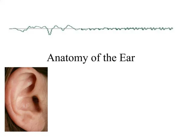
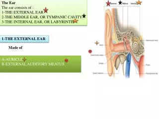
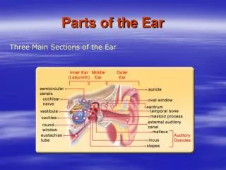
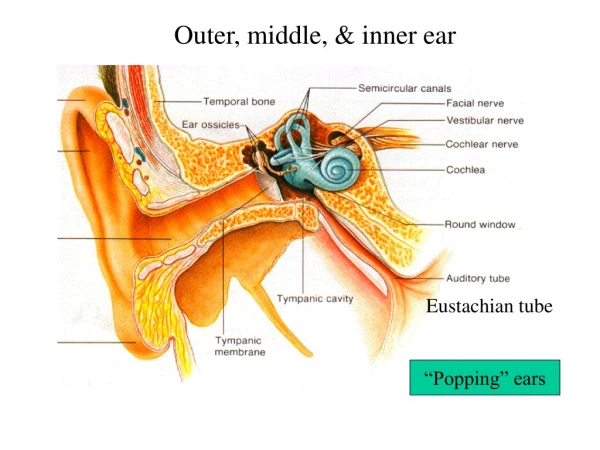
Outer, middle, & inner ear Structures of the Ear Eustachian tube “Popping” ears
Fluid behind tympanum “Tubes in the Ears”
Cranial Nerve VIII Ossicles: incus,malleus, stapes. For transmission & amplification Conduction deafness: ossification of ossicle articulations Attenuation reflex protects cochlea from large vibrations
Scala vestibuli Scala tympani
Endolymph Organ of Corti
Stereocilia with mechanically-gated K+ channels • Open K+ channels • Depolarize • Open Voltage-gated Ca++ channels • Release of NT from synaptic vesicles
Activity of Hair Cells • Depolarization leads to more NT release • Hyperpolarization leads to less NT release
All hair cells nearly identical • Basilar membrane thickens toward the apex
Tonotopy in Cochlea Base Apex
Pitch (frequency) & Intensity • Base .... high pitch (treble) • Apex .... low pitch (bass) • Pitch coded by location of vibrations of Organ of Corti : Which hair cells are stimulated…which set of sensory axons have action potentials • Intensity coded by degree of displacement of stereocilia of hair cells and ultimately the frequency of action potentials in those axons that are active Tonotopy
Pure sine waves Fourier Analysis of Complex Waves Complex wave
Auditory Ranges • Humans: 20- 20,000 Hz; optimal 1000-4000 Hz • Whales: 20 - 100,000 Hz • Bats: 1500 - 100,000 Hz • Frogs: 600 - 3000 Hz • Fish: 20 - 3000 Hz • Crickets: 500 - 5000 Hz
Audiogram Decibel = unit for expressing relative loudness on a log scale “Nerve deafness” cause by damage to hair cells.
Pathway to Temporal Lobe In Brainstem Organ of Corti Hair Cells Cochlear Ganglion = Spiral Ganglion Superior Olive Cochlear Nuclei • VIII cranial nerve • Medial geniculate nucleus of thalamus MG of Thalamus Auditory Cortex
Sound Localization • Low frequency by delay in arrival of soundwave between ears • High frequency by attenuation of intensity • Processed in Superior Olive • Practical Applications? L or R speaker w/ hi and lo frequency tone
Organs of Equilibrium Structures of the Ear Utricle & saccule inside
Semicircular Canal • Angular acceleration • 3 planes
Vestibular Apparatus • Hair cells • NT release dependent upon degree of bending of kinocilium and microvilli • For utricle and saccule: otolith membrane • For ampula of semi-circular canals: cupula • Stereocilia in Endolymph (Hi K+, low Na+)
Additional Topics • Information on cochlear implants • Hearing Aids • Understanding Speaker Frequency Response by Polk Audio’s Marketing Manager • Noise cancelling technology