Download
1 / 41
551 likes | 1.8k Views
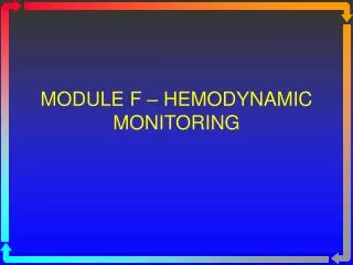
MODULE F – HEMODYNAMIC MONITORING. Topics to be Covered. Cardiac Output Determinants of Stroke Volume Hemodynamic Measurements Pulmonary Artery Catheterization Control of Blood Pressure Heart Failure. Cardiac Measurements.

E N D
Topics to be Covered • Cardiac Output • Determinants of Stroke Volume • Hemodynamic Measurements • Pulmonary Artery Catheterization • Control of Blood Pressure • Heart Failure
Cardiac Measurements • Stroke Volume: Volume of blood ejected from the ventricle with each contraction. • 60-130 mL per beat • Cardiac Output: Volume of blood pumped by the ventricles per minute • CO = SV x HR • Normal Value is 4-8 L/min • 5,040 mL/min = 70 mL x 72/min • Cardiac Output directly influences blood pressure (both HR and SV).
Distribution of Blood Volume • Blood Volume is normally 5 L (varies with age, sex, body size) • 75% is found in the systemic circulation • 60% in the systemic veins • 10% in systemic arteries • 15% is found in the heart • 10% is found in the pulmonary circulation • Usual volume is 75 mL with capacity of 200 mL.
Distribution of Pulmonary Ventilation & Blood Flow • Blood flow is gravity dependent • Sitting/standing position blood goes to the lung bases. • Lying flat, blood flow preferentially will go to posterior lung.
Distribution of Pulmonary Ventilation & Blood Flow • Ventilation is also gravity dependent, BUT… • At FRC, the alveoli are larger at the apices. • Once the FRC is established, the majority of the tidal volume goes to the bases
Distribution of Pulmonary Ventilation & Blood Flow • Net effect is that there is both perfusion and ventilation are greatest at the bases, but not in equal proportions. • West’s Lung Zones • Zone 1 • Least Gravity Dependent. • PA > Pa > PvÞ NO BLOOD FLOW • Zone 2 • Some Gravity Dependency • Pa > PA > PvÞ SOME BLOOD FLOW – AS YOU GO DOWN THE LUNG • Zone 3 • Most Gravity Dependent • Pa > Pv > PA
Changing Body Position to Improve Oxygenation • Unilateral Lung Disease • Examples: Pneumonia only in right lung; fractured ribs with pulmonary contusion only on left. • Bad lung up & Good lung down (GOOD TO GROUND) • Improved ventilation will match greater area of perfusion • Ventilate each lung separately (Independent Lung Ventilation)
Determinants of Cardiac Output • The amount of blood pumped each minute is determined by the number of beats (Heart Rate) and the amount pumped each beat (Stroke Volume). • The Stroke Volume is dependent on three factors: • Preload • Afterload • Contractility • http://www.manbit.com/PAC/chapters/PAC.cfm (look under physiology)
Pulmonary Artery Catheterization • Initially devised by Swan and Ganz. • Allows for view of Left Ventricular function by using extrapolation of right heart measurements. • Catheter is floated into right side of the heart and into a small pulmonary arteriole. • Catheter is then “wedged” and a pressure is measured. • Pulmonary Capillary Wedge Pressure (PCWP) or Pulmonary Occlusion Pressure (POP) • PCWP reflects Left Atrial Pressure (LAP) which reflects Left Ventricular End Diastolic Pressure (LVEDP) which reflects Left Ventricular EndDiastolic Volume (LVEDV)
Preload • Definition • The degree that the myocardial fiber is stretched prior to contraction at end diastole • The more the fiber is stretched, the more it will contract. However, if it is overstretched the amount of contraction goes down. • (Think rubber band) • On right side of heart – Right Ventricular End Diastolic Volume which is reflected by Right Atrial Pressure or Central Venous Pressure (CVP) • On left side, the LVEDV is reflected by the PCWP.
Pulmonary Artery Catheterization • Catheter is inserted into the right side of the heart and advanced into the pulmonary artery. • The physician knows the position of the catheter by watching the waveforms and noting the change in pressures.
http://rnceus.com/hemo/pacath.htm CVP= 2-6 mm Hg
PAP= 25/8 mm Hg (MPAP: 10-20 mm Hg)
Afterload • Definition • The force against which the ventricles must work to pump blood • The ventricular wall tension generated during systole • Determined by: • Volume and viscosity of blood • Vascular resistance • Heart valves
Vascular Resistance • Derivation of Ohm’s Law • The resistance in a circuit is determined by the voltage difference across the circuit and the current flowing through the circuit. • Resistance = D Pressure Flow • Vascular Resistance = D Blood Pressure (mmHg) Cardiac Output (L/min) Where D Blood Pressure is the highest pressure in the circuit minus the lowest pressure in the circuit.
Pulmonary Vascular Resistance (PVR) • Key Components: • Highest Pressure – MPAP • Lowest Pressure – LAP or PCWP • Flow – Cardiac Output • Formula • PVR = (MPAP-PCWP)/CO x 80
Systemic Vascular Resistance (SVR) • Key Components • Highest Pressure – MAP • Lowest Pressure – RAP or CVP • Flow – Cardiac Output • Formula • SVR = (MAP-CVP)/CO x 80
Contractility • Definition • The force generated by the myocardium when the ventricular muscle fibers shorten. • Positive Inotropic effect ( force of contraction) • Negative Inotropic effect (¯ force of contraction) • Contractility is affected by: • Drugs • Oxygen levels within the myocardium • Cardiac muscle damage • Electrolyte imbalances
Heart Failure • Right Heart Failure vs. Cor Pulmonale • Right ventricular hypertrophy • Peripheral edema • Pitting edema, swollen ankles, palpable liver (hepatomegaly), ascites, engorged neck veins (JVD) • Left Heart Failure • Left ventricular hypertrophy • Pulmonary edema and pleural effusions
Types of Invasive Catheters • Arterial Line (A-line) • Inserted into an artery • Central Venous Catheter (CVP) • Inserted into a vein • Pulmonary Artery Catheter (PAP) • Inserted into a vein
Blood Pressure • Low Blood Pressure is hypotension • Prevents the tissues from receiving the O2 and nutrients it needs to survive • High Blood Pressure is hypertension • This strains the heart and over time may lead to heart failure
25/8 (10-20) 4-12 PAP (MPAP) PCWP CVP BP(MAP) 120/80 (80-100) 2-6
Heart/Vascular Pressures • Left Ventricle: 120/0 mm Hg • Arterial Blood Pressure: 120/80 mm Hg • Mean Arterial Pressure: 93 mm Hg (80 - 100) • Arterioles: 30 mm Hg • Capillaries: 20 mm Hg • Veins: 10 mm Hg • Right Atrium (CVP): 2-6 mm Hg (4-12 cm H20) • Right Ventricle 25/0 mm Hg • Pulmonary Artery 25/8 mm Hg • Mean Pulmonary Artery Pressure: 14-15 mm Hg (10-20 cm H2O) • Pulmonary Capillary Wedge Pressure (PCWP): 4-12 mm Hg • Pulmonary capillaries 12 mm Hg • Pulmonary veins 8-10 mm Hg • Left Atrium 5 mm Hg • Left Ventricle - 120/0 Page 7 of Formulae
Factors that Control Blood Pressure • Heart • HR and Stroke Volume, BP • ¯ HR and Stroke Volume, ¯ BP • Blood • Hypervolemia: Increased BP • Hypovolemia: Reduced BP • Blood Vessels • Vasoconstriction: Increased BP • Vasodilation: Reduced BP
Mean Blood Pressure • Mean = 2 (diastolic pressure) + systolic pressure 3 • Mean arterial blood pressure is 90–100 mm Hg • Mean pulmonary artery pressure is 9-18 mm Hg
Indexed Values • Relates values to body size • Allows for better correlation between patients • Calculated by using Dubois nomogram • Appendix IV, p 513 • Cardiac Index • CO/BSA • Stroke Volume Index • SV/BSA • Pulmonary & Systemic Vascular Resistance Index • Divide by CI instead of CO