Download

1 / 55
590 likes | 758 Views
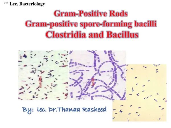
GRAM POSITIVE BACILLI. Clinically important Gram positive bacilli. Spore forming Bacillus Clostridium Non spore forming 1.Corynebacterium 2.Listeria 3.Lactobacillus. Bacilli w/ branching filaments 1.Actinomyces 2.Nocardia. 1.BACILLUS. Bacillus anthracis Human pathogen

E N D
Clinically important Gram positive bacilli Spore forming • Bacillus • Clostridium Non spore forming 1.Corynebacterium 2.Listeria 3.Lactobacillus Bacilli w/ branching filaments 1.Actinomyces 2.Nocardia
1.BACILLUS • Bacillus anthracis • Human pathogen • Isolation also considered to be clinically significant • Zoonosis • Bacillus cereus • Environmental organism • Contaminates food • Common cause of food poisoning • Bacillus stearothermophilus • Tolerates very high temperatures • Used for quality control of autoclaves
a.Bacillus anthracis • Large bacilli of 1-3m • Historical importance • Single or paired in clinical isolates • In vitro – prominent capsule • Highly resistant spores
Anthrax Pathogenesis and clinical presentations Cutaneous anthrax About 20% mortality Virulence factors Capsule (antiphagocytic) Toxin (oedema & death) Inhalation anthrax High mortality Gastrointestinal anthrax High mortality
Anthrax - Diagnosis • Specimen • Aspirate or swab from cutaneous lesion • Blood culture • Sputum • Laboratory investigation • Gram stain • Culture • Identification of isolate
Anthrax – treatment and prevention • Penicillin • (Tetracycline /chloramphenicol) • Erythromycine,Clindamicine • Prevention • Vaccination of animal herds • Proper disposal of carcasses • Active immunisation with live attenuated bacilli
b.Bacillus cereus • Large, motile, saprophytic bacillus • Heat resistant spores • Pre formed heat and acid stable toxin (Emetic syndrome) • Heat labile enterotoxin (Diarrhoeal disease) • Lab diagnosis – Demonstation of large number of bacilli in food
Bacillus cereus clinical presentation Gastroenteritis EMETIC FORM DIARRHOEALFORM Incubation period > 6 hours Diarrhoea Lasts 20-36 hours Incubation period < 6 hours Severe vomiting Lasts 8-10 hours
CLOSTRIDIUM(ANAROBES) • Anaerobic • Sporing • Gram positive • Diameter of the spore is larger than the cell resemble a spindle • Clostridium is derived from Kloster meaning spindle
Spores Pleomrhic (elongated, spindle) Most are obligate anaerobes produce neuro histo toxins
Saprophytes - Most • Some are opportunists - tetanus/gas gangrene/food poisoning • Cl. perfringens - commensal of the intestine • Cl. sporogenes - -do- • Can invade the intestine after the death
CLASSIFICATION BASED ON THE TYPE OF DISEASE PRODUCED • A . Tetanus Cl. tetani - Present in soil • B. Gas gangrene • Established Cl. perfringens ‘gut’ organism Cl. septicum Cl. novyi - Less pathogenic Cl. histolyticum Cl. fallax - Doubtful Cl. bifermentans Cl. sporogenes
C. Food poisoning 1. Gastroenterritis - Cl perfringens Type A 2. Botulism - Cl. botulinum/ Soil 3. Pig-belCl. perfringens type C • D. Acute colitis - Cl. difficile / gut’ organism (pseudomembranous colitis) • Commonest cause of ‘nosocomial’ diarrhoea
Dead tissue, blood clots, foreign matter aerobic organisms In an injury DEVELOP ANAEROBIC CONDITION (Exogenous infection) Germination of spores Gas gangrene oedema, necrosis, gas production, toxaemia, myositis Crepitus
C Perfringens C histolyticum C septicum C novyii C Perfringens Alpha toxin (lecithinase)
TETANUS Cause tetanus in both man and animals disease which effect the nervous system of the host. • Agricultural workers and gardeners and are more prone because the spores are present in the soil. • At birth under unhygienic conditions baby’s can get – tetanus neonatorum.
Soil/Intestine/Vagina • Drum stick appearance • Motile with peritrichous flagella • Obligatory anaerobes • Grow on Robertson’s cooked medium
All types produce the same toxin • C. tetani – 10 types based on the H antigens • (CP – 5 types based on the type of toxins, alpha, beta, epsilon, iota).
Susceptibility - Some strains can withstand boiling for 3hrs/dry heat 1600C for 1hr. but all will destroy at 1210C/15 min.
COMMON FEATURES FOR BOTH CT AND CP • All CP’s produce alpha toxin • All CT’s produce same exotoxin – plasmid mediated • However, CP’s got enterotoxins. • Exotoxin of CT has got two components • .Tetanolysin – both heat and O2 labile – may act as a leucocidin • .Tetanospasmin – heat labile, but O2 stable (Therefore, can you give an edvantage ? will not get destroyed in the blood).
Spores germinate -------toxin-----motor nerve endings--------along the motor neurones of the peripheral nerve to the anterior horn cells------local tetanus (in the proximity of the wound). • Ascending tetanus – when toxins spreads upwards along the spinal cord towards C.N.S. Gives generalized spasms. • Descending tetanus – when toxin is given IV , spasms will appear in the muscles of the head, neck and spreads downwards.
Clinical symptoms • Early symptom is trismus (lock jaw) – spasms of the masseter muscle • difficulty in opening of the mouth and masticating • rigidity spreads to muscles of the face, neck and truck • risus sardonicus – contraction of the frontails and muscles at the angle of the mouth • back is usually slightly curved (Opisthonotus ?) • Insevere cases violent spasms will last for few seconds to 3-4 mins. • If convulsions appear soon after the initial symptoms, it is very serious. • The spasms gradually intensify and patient may die of • .exhaustion, b. asphyxia or aspiration peumonia • - If local tetanus after a wound at the neck, you might think of tuberculous meningitis (irritation and paralysis is common). • What happens • Toxin acts at the synaptic junction – prevent the synthesis of acetylcholine. Thus, prevents synaptic transmission.
Toxins • Tetanolysin - heat and oxygen labile/lyse RBC/ • Tetanospasmin - heat and oxygen stable/highly lethal (for mice 0.0000001 mg) dies within 1 - 2 days get easily neutralize with antitoxin
GABA GLYCINE
Clostridial food poisonng • C. perfringens • Carriers for food poisoning strains • Survival of heat resistant spores in bulk meals • Sporulation in gut - Short IP and watery diarrhoea for 24-48 hours • Beta toxin production in C. prerfringens type C – Necrotizing enteritis(Pig bell)
BOTULISM Sausage 8 toxins (A-G) Food borne botulism (IP 1-2 days) Infant botulism Wound botulism (IP > 4 days) Diagnosis Isolation of organism in food/faeces Detection of toxin in faeces / serum
Produces Botulism World wide distribution Found in soil and occasionally in animal feces Sporese are highly heat resistant ,withstand 100C for 3-5 hrs. Heat resistance is reduced by acid pH or high salt concentrations Toxin Released during growth and autolysis of bacteria. It is found in 7 antigenic varieties.A-G The principle cause for human disease A,B,E/F
A,B - Variety of foods E - Fish products C - Limberneck in birds D - botulism in mammals Toxin is neurotoxic protein Destroyed by heating at 100C for 20 mins. Action :Block release of Acetylecholine at synapses and NMJ causing flaccid paralysis. Pathogenecity Illness is not an infection. Botulism is an intoxication resulting from the ingestion of food in which C.botulinum has produced toxin.
PSEUDOMEMBRANOUS COLITIS Virulence factors Enterotoxin (Toxin A) Cytotoxin (Toxin B) Management Discontinue antibiotics Ampi/Tetra/Clinda Oral metronidazole Oral vancomycin Diagnosis Clinical suspicion Culture of faeces Detection of toxin
CORYNEBACTERIA(AEROBES) • Causes localized inflammation (pseudomembrane, greyish white exudate ) and generalized toxaemia • Prevalent in baby’s after 3-6 months (that’s why DPT is given at 3, 5, 7 months, boosters at 18 months and at school entry), very high in young children
Morphology • Gram/+ve/palisade/Chineseletter arrangement • Irregular swellings at one end -club shaped. • Corynebacteria tend to pleomorphism in microscopic and colonial morphology.
On blood agar Small granular & gray with irregular edges and may have small zones of hemolysis. • Grow aerobically on ordinary media
a. Corynebacterium diphtheriae Normal flora of nasopharynx in about 10% • Diphtheria caused when infected by lysogenic bacteriophage b. Diptheroids • Normal flora of skin • Usual contaminants of samples • Can cause disease in ‘compromised’ host C. ulcerans C. haemolyticum C. jeikeium
Rare in developed countries/ third world countries • Nose, Nasopharynx, skin aerobic, facultatively anaerobic • Nasal carriers are very dangerous
Loeffler's serum slope Blood telurite agar (black colonies) • Morphological differences • Three biotypes Gravis (severe) Inter-medius (intermediate) Mitis (mild)
Epidemiology • It is rare in developing countries, a disease of the third world countries. Still highly prevalent in the former Soviet Union. • Spread through droplets.
Types of Diphtheria • Faucial • Laryngeal • Nasal • Conjunctival • Vulvovaginal • Otitic • Cutaneous around the mouth and the nose
Effect of toxins 1. Local 2. General Toxaemia and acts on the myocardium and on motor nerves and adrenals Complications a, pseudomembrane may extend to larynx and cause obstruction b.myocarditis /Polyneuropathy • Degenerative changes in the liver adrenals, kidney's
Pathology • Toxin is absorbed in the mucus membrane and causes • destruction of epethelium and causes a superficial • inflammatory respons. • Necrotic epethelium becomes embeded in exuding fibrin and red and white cells, with bacteria- • Grayish pseudomembrane is formed over the tonsilas • and pharynx and larynx.
Removal of pseudomembrane - capillary damage and • bleeding.. • Regional lymphadynopathy with marked edema of the • neck within the membrane bacilli produce toxin. • This results in distant toxic damage paranchymatous • degeneration fatty infiltration & necrosis in heart • muscle liver kidney & adrenals.
How to identify the immune persons Shick test – suitably diluted stabilized toxin intradermally, localized erythema (1-3cm) in 2-4 days, means no or little antibodies
Diagnosis • Direct smear - Albert's stain • Culture - Loffler's serum slope/blood agar/blood telurite agar Check the toxigenicity • Animal inoculation Death within 96 hrs Guinea pigs/rabbits