Download
1 / 16
190 likes | 610 Views
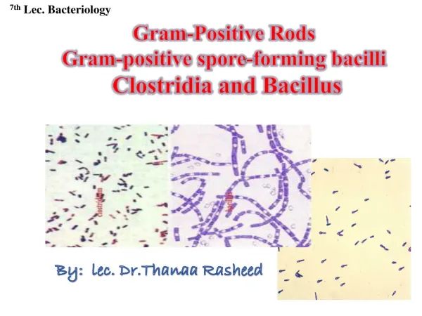
Gram-Positive Bacilli Part One. MLAB 2434: Microbiology Keri Brophy -Martinez. General Characteristics: Bacillus. Habitat Thermal springs, fresh and salt water, soil, on plants Aerobic Spore-formers Resistant to radiation, heat, disinfections, and desiccation

E N D
Gram-Positive BacilliPart One MLAB 2434: Microbiology Keri Brophy-Martinez
General Characteristics: Bacillus • Habitat • Thermal springs, fresh and salt water, soil, on plants • Aerobic • Spore-formers • Resistant to radiation, heat, disinfections, and desiccation • Mostly contaminants in clinical specimens • EXCEPTION: Bacillus anthracis
More on Spores • Produced when the bacteria gets stressed • Drying • Temperature extremes • Aid in organism’s survival • Heat shock induces spores • Temperature raised to 56o • Gram stain • Appear as clear areas within the bacterial cell
Significant Bacillus Species • Bacillus anthracis • Agent of anthrax, a disease in livestock • Humans acquire infection by contamination of wound or ingestion or inhalation of spores • Bacillus cereus • Causes food poisoning • An opportunist • Bacillus subtilis • Common laboratory contaminant • Used in sterility testing
Bacillus anthracis • Cutaneous anthrax • “Malignant pustule” (also called “black escher”) • Woolsorter’s disease/ Rag-pickers disease • Organisms gain access through cuts; localized infection • Majority of cases in the world are cutaneous • Inhalation anthrax • Acquired through inhalation of spores • May result in respiratory distress and death • Gastrointestinal • Acquired by ingestion of contaminated raw meat • Usually fatal
Bacillus anthracis: Clinical Infections in Humans • Cutaneous anthrax
Laboratory Diagnosis • Media • Most species grow well on SBA • Most species beta-hemolytic, exceptB. anthracis • No growth on MacConkey • Fast growers • Colony characteristics vary • Catalase positive
Laboratory Diagnosis • Goal in identification is to RULE OUT B. anthracis • If B. anthracis is suspect, MUST work under safety hood • Other Bacillus, identified to genus level ONLY
Identification of Bacillus anthracis • Microscopic morphology • Large, square-ended gram-positive rods • Bamboo appearance • Spores may be absent in patient smears
Identification of Bacillus anthracis • Colony Morphology • Nonhemolytic on blood agar; raised, large, grayish white, irregular, fingerlike edges • “Medusa head” or “beaten egg whites”
Bacillus cereus • Food poisoning • Diarrheal syndrome • Associated with meat, poultry, and soups • Incubation period of 8 to16 hours • Fever uncommon • Resolves within 24 hours • Emetic form • Associated with fried rice • Abdominal cramps and vomiting • Incubation period of 1 to 5 hours • Resolves in 9 hours
Bacillus cereus • Local infections • Postsurgical/traumatic wounds • Burns • Eye infections • Rare conditions • Meningitis • Bacteremia • Endocarditis • Osteomyelitis
Bacillus subtilis • Found in the environment • Common laboratory and hospital contaminant • Used as a QC agent for sterilization procedures
References • http://www.atsu.edu/faculty/chamberlain/golden2000/case5.htm • http://chesschumpion.blogspot.com/2007/03/time-to-put-on-your-thinking-caps.html • http://en.wikipedia.org/wiki/File:Bacillus_subtilis_Gram.jpg • https://labs.uhstx.com/clinical_int/dols/appb.htmlhttp://www.iccb.state.il.us/pt3/mod/science/mod_bio111/mod10/p4.html • http://www.flickr.com/photos/microbeworld/sets/72157625392265538/detail/http://www.uaz.edu.mx/histo/pathology/ed/ch_9b/c9b_clue.htm • Kiser, K. M., Payne, W. C., & Taff, T. A. (2011). Clinical Laboratory Microbiology: A Practical Approach . Upper Saddle River, NJ: Pearson Education. • Mahon, C. R., Lehman, D. C., & Manuselis, G. (2011). Textbook of Diagnostic Microbiology (4th ed.). Maryland Heights, MO: Saunders.