Download
1 / 33
400 likes | 751 Views
Malignant Mesothelioma in Effusions and Fine Needle Aspirates. Armando C. Filie, M.D. National Cancer Institute. No relationship exists that represents a possible conflict of interest with respect to the content of this presentation. OBJECTIVES. Objectives

E N D
Malignant Mesothelioma in Effusions and Fine Needle Aspirates Armando C. Filie, M.D. National Cancer Institute No relationship exists that represents a possible conflict of interest with respect to the content of this presentation
OBJECTIVES • Objectives • Recognize the cytological features of malignant mesothelioma (mesothelioma) in effusion samples • Recognize the cytological features of fine needle aspirates of mesothelioma • Recognize the cytological features of major lesions in the differential diagnosis of mesothelioma • Familiarize with current ancillary studies in the diagnosis of mesothelioma
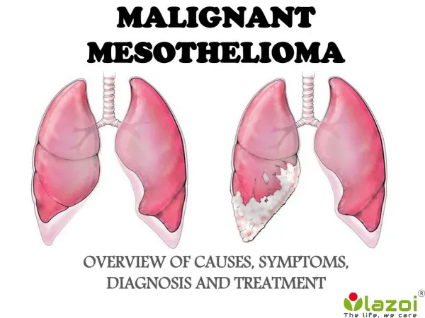
BLANK SLIDE Mesothelioma • Malignant neoplasm of pleura, peritoneal cavity and pericardium • Incidence of 2,500 cases/year (pleural) • Clinical Findings • age and presentation: males, 6th-8th decade, unilateral • pathogenesis: asbestos exposure (latency of 20-50 years), ?simian vacuolating virus (SV40) • imaging findings: CT scan [pleural masse(s)], invasion by magnetic resonance imaging (MRI) • Diagnosis: clinical history + imaging findings + cytology(?)/biopsy
Mesothelioma • Prognosis and Treatment • poor prognosis • treatment: surgery (most effective), chemotherapy, radiotherapy (localized recurrences), combine therapy • Histologic Types • epithelioid (epithelial): up to 17 subtypes (deciduoid, clear cell, small cell, signet ring) • sarcomatoid: 8 subtypes (fibrosarcomatous, lymphohistiocytoid, MFH-like) • biphasic (mixed) • desmoplastic
Mesothelioma • Cytological Features in Effusions • sample preparation: smear, cytocentrifugation, thin layer, cell block (immunostains) • stains: Diff-Quik, Papanicolaou
Mesothelioma • Cytological Features in Effusions • patterns: epithelioid (malignant epithelial), sarcomatous (sarcomatoid), anaplastic, biphasic • sarcomatoid mesothelioma differential diagnosis: spindle cell sarcomas • biphasic mesothelioma differential diagnosis: carcinomas (renal cell carcinoma) • anaplastic mesothelioma differential diagnosis: pleomorphic sarcomas • epithelioid mesothelioma: most frequent pattern, associated with effusion more frequently than other patterns.
Mesothelioma in Effusions Cytological Features of Epithelioid Mesothelioma • cellular sample • one cell population • clusters (scalloped border) • cell-in-cell formations • intercellular spaces (“windows”) • two-tone cytoplasm • surface blebs • variable N/C ratio • multinucleation • macronucleoli
Mesothelioma in Effusions Cytological Features of Epithelioid Mesothelioma
Mesothelioma in Effusions Cytological Features of Epithelioid Mesothelioma
Mesothelioma in Effusions Differential Diagnosis • Metastatic carcinoma: adenocarcinomas (lung, breast, gynecologic tract, gastrointestinal tract), may be the first manifestation of an occult primary • Hematologic neoplasms: B-cell lymphomas (diffuse large B-cell), T-cell lymphomas (anaplastic large cell), plasma cell neoplasms, primary effusion lymphoma (PEL) • Melanoma: may be the first manifestation of disease • Others: squamous cell carcinoma, mesothelial cell lesions
Mesothelioma in Effusions Cytological Features of Metastatic Adenocarcinoma • cellular sample • two cell population • clusters (smooth border) • cell-in-cell formations • high N/C ratio • multinucleation • macronucleoli • irregular nuclear contours • delicate/dense cytoplasm • vacuole(s) displacing the nucleus
Mesothelioma in Effusions Cytological Features of Metastatic Adenocarcinoma
Mesothelioma in Effusions Cytological Features of Metastatic Melanoma • cellular sample • two cell population (?) • aggregates • cell-in-cell formations • low N/C ratio • multinucleation • macronucleoli • intranuclear cytoplasmic inclusions • melanin pigment • vacuoles
Mesothelioma in Effusions Cytological Features of Metastatic Melanoma
Mesothelioma in Effusions Cytological Features of PEL • cellular sample • two cell population • variable N/C ratio • multinucleation • macronucleoli • dense basophilic cytoplasm
Mesothelioma in Effusions Cytological Features of PEL
Mesothelioma in Fine Needle Aspirates • Image-guided fine needle aspiration (FNA) may be used for the initial diagnosis of mesothelioma • 4% needle tract seeding for core-needle biopsy with sensitivity of 86% (pleural) • FNA of metastatic mesothelioma (rare): scalp, thyroid, cervical lymph node, axillary lymph node, subcutaneous nodules, breast, liver • metastasis may be the first indication of mesothelioma • inclusions of benign mesothelial cells in lymph nodes • Mesothelial cell lesions of pleura: solitary fibrous tumor (most benign, rare malignant), nodular pleural plaque, adenomatoid tumor, simple mesothelial cyst, multicystic mesothelioma, well-differentiated papillary mesothelioma, localized malignant mesothelioma
Mesothelioma in Fine Needle Aspirates Cytological Features of Mesothelioma in FNAs • cellular aspirate • clusters and flat sheets • papillary groups (core) • acinar/tubular groups • single cells • intercellular spaces • round/polygonal shape • spindle cells (sarcomatoid, biphasic) • small cytoplasmic vacuoles • multinucleation
Mesothelioma in Fine Needle Aspirates Cytological Features of Mesothelioma in FNAs
Mesothelioma in Fine Needle Aspirates Differential Diagnosis • Epithelioid: carcinoma - lung (adenocarcinoma and bronchoalveolar carcinoma [BAC]), ovary and peritoneal serous carcinoma; mesothelial cell lesions; thymoma; epithelioid sarcomas, reactive mesothelial proliferations • Sarcomatoid: mesothelial cell lesions, desmoid tumor, schwannoma, spindle cell sarcomas • Biphasic: thymoma, synovial sarcoma, desmoplastic small round cell tumor, pleuropulmonary blastoma • Anaplastic: pleomorphic sarcomas
Mesothelioma in Fine Needle Aspirates Cytological Features of Lung BAC in FNAs • monolayer sheets • papillae • single cells • round nuclei • nuclear grooves and pseudoinclusions • nuclear crowding/overlapping • pleomorphic cells • mucin (mucinous)
Mesothelioma in Fine Needle Aspirates Cytological Features of Lung BAC in FNAs
Mesothelioma (Ancillary Studies) • Histochemical stains: mucin (Alcian blue, mucicarmin) • Electron microscopy: long microvilli (meso), short (adeno) • FISH: detection of chromosomal alterations • Hyaluronic acid levels in effusion samples • Immunocytochemistry: most commonly used • may be applied to cytocentrifuged samples, smears, thin layer samples, cell blocks (preferred) • panel of mesothelial cell and adenocarcinoma markers: 2 meso and 2 adeno markers or 1/2 meso and 3 adeno markers • other markers: hematopoietic markers, melanoma markers, site “specific” markers (TTF-1, PSA, PAP, CDX-2, GCDFP-15, thyroglobulin)
Mesothelioma (Ancillary Studies) Mesothelial cell (Mesothelioma) Markers • calretinin: neuron-specific calcium binding protein (neural tissues and a few other cell types like mesothelial cells) • cytokeratin 5/6: intermediate filament (mainly keratinized and non-keratinized squamous cell carcinoma) • Others: HBME-1, WT1,Mesothelin, Podoplanin calretinin
Mesothelioma (Ancillary Studies) Mesothelial cell (Mesothelioma) Markers HBME-1 CK 5/6
Mesothelioma (Ancillary Studies) Adenocarcinoma Markers • B72.3: antibody detects a tumor associated protein • Ber-EP4: antibody against epithelial adhesion molecule • CA19.9: antibody against Lewisa blood group antigen • Others: mCEA, CD15, MOC-31 B72.3
Mesothelioma (Ancillary Studies) Adenocarcinoma Markers B72.3 CA19.9 Ber-EP4
Mesothelioma (Ancillary Studies) • Melanoma markers: HMB45, Mart-1, KBA62, S100 • Hematopoietic markers: LCA, L26, CD38, HHV8 • Others: TTF-1, PSA and PAP, GCDFP-15, thyroglobulin HMB45 Mart-1
Mesothelioma (Ancillary Studies) Hematopoietic and other markers HHV8 TTF-1 CK 7
Mesothelioma (Ancillary Studies) Molecular Tests • Gene expression (quantitative RT-PCR) • Proteomics: protein complement of the genome (serum - early cancer diagnosis), potential in cytopathology • Surface enhanced laser desorption/ionization time of flight (SELDI-TOF): protein profile in cytology samples (Fetsch et al, 2002) • Initial set: 5 renal cell carcinomas, 9 metastatic melanomas, 6 reactive effusions • Unknown set: 4 renal cell carcinomas, 8 metastatic melanomas, 3 reactive effusions
Mesothelioma (Ancillary Studies) Molecular Tests • SELDI-TOF in FNAs and fluid samples of 8 MM, 4 RCC, 3 reactive effusions
Mesothelioma in Effusions and FNAs SUMMARY • Cytological features of mesothelioma in effusions and FNAs overlap with those seen in other benign and malignant lesions (adenocarcinoma) • Some cytologic features of mesothelioma are not often present in cytology samples of lesions that should be considered in the differential diagnosis • Ancillary studies are important in supporting the diagnosis of mesothelioma (immunocytochemistry, electron microscopy) • Diagnosis of mesothelioma has prognostic, treatment and legal implications