Download
1 / 2
20 likes | 231 Views
Case Report : Malignant Peritoneal Mesothelioma Jung Woo Park.MD, Sung Ook Hwang.MD, En Seop Song.MD, Young Chae Chu.MD*, Woo Young Lee.MD, From the Department of Obstetrics & Gynecology, Department of Pathology*, Inha University Hospital, Incheon, Republic of Korea .

E N D
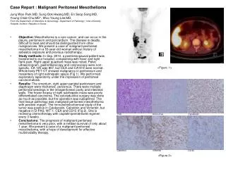
Case Report : Malignant Peritoneal Mesothelioma Jung Woo Park.MD, Sung Ook Hwang.MD, En Seop Song.MD, Young Chae Chu.MD*, Woo Young Lee.MD, From the Department of Obstetrics & Gynecology, Department of Pathology*, Inha University Hospital, Incheon, Republic of Korea. Objective: Mesothelioma is a rare cancer, and can occur in the pleura, peritoneum and pericardium. The disease is deadly, difficult to treat and should be distinguished from other malignancies. We present a case of malignant peritoneal mesothelioma in a 53-year-old woman without history of asbestos exposure and previous radiotherapy. Study methods: In Sep. 2010, a postmenopausal patient was transferred to our hospital, complaining with fever and right flank pain. Right upper quadrant mass was noted. Pelvic ultrasonogram, gastrofiberscopy and colonoscopy were non-specific. CA 125 was 867, but CEA and CA19-9 were normal. Whole body PET-CT showed malignancy in peritoneum and mesentery of right subhepatic space (Fig 1). We performed exploratory laparotomy under the impression of peritoneal carcinomatosis. Results: The omentum, right upper parietal peritoneum and diaphragm were thickened, cancerous. There were multiple peritoneal seedings in the intraperitoneal cavity and intestinal walls. The frozen biopsy of right subhepatic mass was poorly differentiated carcinoma. The cytoreductive surgery was done as much as possible, but the operation was suboptimal. The final tissue pathology was malignant peritoneal mesothelioma with positive margin. The immunohistochemical study of the tumor was positive in Cytokeratin, Calretinin and Vimentin, but negative in D-PAS, WT-1, CEA and CD15 (Fig 2). She is receiving chemotherapy with cisplatin/pemetrexed regimen every 3 weeks . Conclusions: The prognosis of malignant peritoneal mesothelioma is very poor, with a median survival of only about 1 year. We present a case of a malignant peritoneal mesothelioma, with a hope of development for effective multimodality therapy. <Figure. 1> <Figure 2> PAS Alcian blue Cytokeratin Calretinin Vimentin D2-40
Case Report : Malignant Peritoneal Mesothelioma Jung Woo Park.MD, Sung Ook Hwang.MD, En Seop Song.MD, Young Chae Chu.MD*, Woo Young Lee.MD, From the Department of Obstetrics & Gynecology, Department of Pathology*, Inha University Hospital, Incheon, Republic of Korea. Objective: Mesothelioma is a rare cancer, and can occur in the pleura, peritoneum and pericardium. The disease is deadly, difficult to treat and should be distinguished from other malignancies. We present a case of malignant peritoneal mesothelioma in a 53-year-old woman without history of asbestos exposure and previous radiotherapy. Study methods: In Sep. 2010, a postmenopausal patient was transferred to our hospital, complaining with fever and right flank pain. Right upper quadrant mass was noted. Pelvic ultrasonogram, gastrofiberscopy and colonoscopy were non-specific. CA 125 was 867, but CEA and CA19-9 were normal. Whole body PET-CT showed malignancy in peritoneum and mesentery of right subhepatic space (Fig 1). We performed exploratory laparotomy under the impression of peritoneal carcinomatosis. Results: The omentum, right upper parietal peritoneum and diaphragm were thickened, cancerous. There were multiple peritoneal seedings in the intraperitoneal cavity and intestinal walls. The frozen biopsy of right subhepatic mass was poorly differentiated carcinoma. The cytoreductive surgery was done as much as possible, but the operation was suboptimal. The final tissue pathology was malignant peritoneal mesothelioma with positive margin. The immunohistochemical study of the tumor was positive in Cytokeratin, Calretinin and Vimentin, but negative in D-PAS, WT-1, CEA and CD15 (Fig 2). She is receiving chemotherapy with cisplatin/pemetrexed regimen every 3 weeks . Conclusions: The prognosis of malignant peritoneal mesothelioma is very poor, with a median survival of only about 1 year. We present a case of a malignant peritoneal mesothelioma, with a hope of development for effective multimodality therapy. <Figure. 1> <Figure 2> PAS Alcian blue Cytokeratin Calretinin Vimentin D2-40