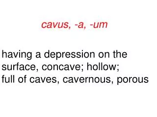
Cavus Foot
Cavus Foot. N. Craig Stone M.D. F.R.C.S.(C) Discipline of Orthopedic Surgery Sept 29, 2003. Cavus Foot Introduction. Definition Anatomy and Pathomechanics Etiology and differential diagnosis Evaluation Clinical and radiographic Treatment. Cavus Foot Definition.


Cavus Foot
E N D
Presentation Transcript
Cavus Foot N. Craig Stone M.D. F.R.C.S.(C) Discipline of Orthopedic Surgery Sept 29, 2003
Cavus FootIntroduction • Definition • Anatomy and Pathomechanics • Etiology and differential diagnosis • Evaluation • Clinical and radiographic • Treatment
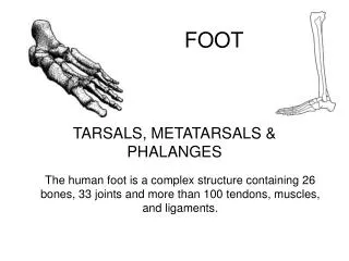
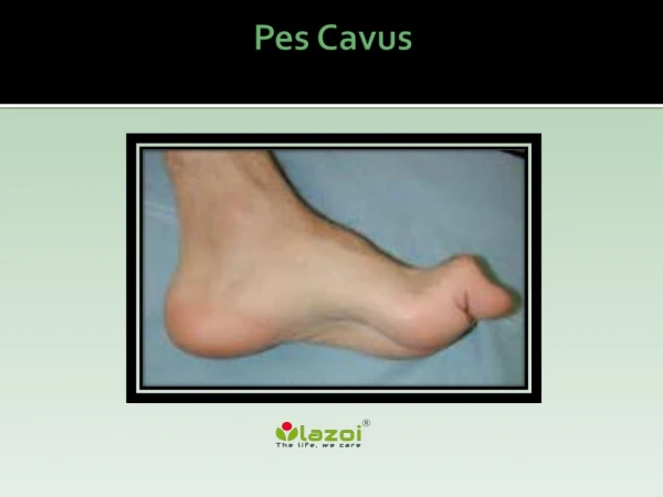
Cavus FootDefinition • Abnormal elevation of the medial arch in weight bearing • Fore foot equinus relative to hindfoot • ?what’s normal/abnormal
Normal Anatomy and Biomechanics • Forefoot deformity and the windlass mechanism of the plantar fascia causative • Plantar fascia • Calcaneal tuberosity – Transverse metatarsal lig – slips to base of prox phalanx • Medial and central portions strongest • Stabilizes arch and inverts (with tib post) the hindfoot
Anatomy and Biomechanics • Chopart’s joint supple when hindfoot everted • Heel strike – hindfoot inverted • Midstance – hindfoot everted • Shock absorption – now hindfoot supple • Toe off
Anatomy and Biomechanics • Toe off • Toes dorsiflex • Tib post fires • All to lock hindfoot • Gives a rigid, long lever for triceps surae
Pathomechanics • Foot musculature unbalanced • Usually intrinsic muscle weakness • Lumbrical weakness allows EDL to hyperextend the MCP’s and FDL to flex the PIP and DIP’s • Exaggeration of the windlass mechanism
Pathomechanics • Same applied to EHL and FHL • 1st ray more mobile – makes it worse, forefoot supinates and may become fixed • Secondary hindfoot varus • Tripod effect
Pathomechanics • So why does it hurt? • Inverted hindfoot loses shock absorption ability • Recurrent ankle sprains • Tripod effects (less surface area) • Clawing of toes
Etiology • CNS • Spinal • Peripheral Nerves • Other • Idiopathic
Etiology - CNS • CP esp. hemiplegia • Spastic tib post • Friedreich’s Ataxia (A. Recessive chrom 9) • Triad – ataxia, downgoing Babinski, areflexia
Etiology - Spinal • Myelodysplasia • Syringomyelia • Polio • Spinal cord tumors • Tethered cord • Guillain-Barre syndrome
Etiology – Peripheral Nerves • Hereditary Sensorimotor Neuropathy (HSMN) • Charcot Marie Tooth
Etiology - Other • Traumatic Isolated Tendon Injuries • Partial Sciatic Nerve injury • Volkman’s Contracture
Etiology - Idiopathic • 20-50% of cases - mostly bilateral
Clinical Evaluation • History • Other neuro symptoms • Ulcers, numbness, bowel, bladder, Dev. Delay • Family History • Ankle Instability • Metatarsalgia
Clinical Evaluation • Physical • Dysraphism • Neuro exam • Coleman Block Test
Radiographic Assessment • Standing AP and Lateral of Foot and Ankle • Assess angles (severity) and any evidence of degenerative change • Spinal Imaging as required
A – Meary’s Angle N = 0 – 5 Degrees B – Calcaneal Pitch Angle N = 30 degrees C – Hibbs Angle N = <45 degrees D – Weight Bearing Tibioplantar Angle N = 90 degrees
Management • Blah, Blah, Blah • Orthotics • For mild, non progressive deformity • Lateral forefoot and hindfoot posting • Large toe box shoes
Management • Surgical • Treat underlying problem • Must decide if hindfoot is supple • Everyone gets a plantar fascial release • Fixed – Supple is often subjective • Combination of procedures
Management • Hindfoot supple • Toe deformity correction • Girdlestone-Taylor • Forefoot correction • Metatarsal osteotomies • Midfoot osteotomy • Is there any role for a Jones procedure?
Management • Hindfoot supple • Tendon Transfers • If identifiable muscle imbalance • Split Tib post to peroneus brevis • Peroneus longus to brevis • Be careful in progressive disease
ManagementRigid Hindfoot • If deformity severe or Degenerative Changes exist • Triple Arthrodesis
Summary • Rare problem • Know causes and clinical assessment • Principles of treatment