Download
1 / 48
480 likes | 651 Views
WINDSOR UNIVERSITY SCHOOL OF MEDICINE St.Kitts. Dr. SREEKANTH THOTA. DEPARTMENT OF ANATOMY. UPPER LIMB. FOREARM . FOREARM. The forearm is the part of the upper limb that extends between the elbow joint and the wrist joint.

E N D
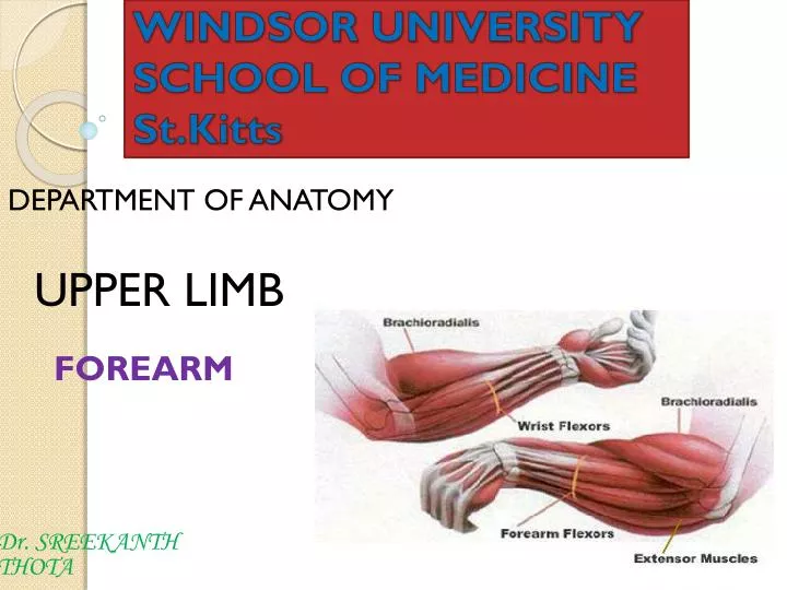
WINDSOR UNIVERSITYSCHOOL OF MEDICINESt.Kitts Dr. SREEKANTH THOTA DEPARTMENT OF ANATOMY UPPER LIMB FOREARM
FOREARM • The forearm is the part of the upper limb that extends between the elbow joint and the wrist joint. • As in the arm, the forearm is divided into anterior and posterior compartments
Compartments of forearm • Muscles in the anterior compartment of the forearm flex the wrist and digits and pronate the hand. • Muscles in the posterior compartment extend the wrist and digits and supinate the hand.
ANTERIOR COMPARTMENT OF THE FOREARM • Muscles in the anterior (flexor) compartment of the forearm occur in three layers: • 1. Superficial • 2. Intermediate • 3.Deep • All muscles in the anterior compartment of the forearm are innervated by the median nerve, except for the flexor carpiulnarismuscle and the medial half of the flexor digitorumprofundusmuscle, which are innervated by the ulnar nerve.
Superficial layer • All four muscles in the superficial layer-1.flexor carpiulnaris • 2.palmaris longus • 3.flexor carpiradialis • 4. pronatorteres- • All four muscles have a common origin from the medial epicondyleof the humerus, and, except for pronatorteres, extend distally from the forearm into the hand
Intermediate layer • Flexor digitorumsuperficialis
Deep layer • 1.flexor digitorum profundus • 2.flexor pollicis longus • 3.pronator quadratus
Arteries of the anterior compartment of the forearm. • At the apex of the cubitalfossa, Brachial artery divides into its two major branches, the radial and ulnararteries.
Radial artery • In the distal forearm, the radial artery can be located using the flexor carpiradialis muscle as a landmark. • Radial artery lies immediately lateral to the large tendon of the flexor carpiradialis muscle.
Branches of the radial artery • 1. Radial recurrent artery - arises just after the radial artery comes off the brachial artery. • 2. Superficial palmar branchenters the hand by passing through, or superficial to, the thenar muscles at the base of the thumb
Radial pulse • The radial pulse can be felt by gently palpating the radial artery against the underlying muscle and bone.
Ulnar artery • The ulnar artery is larger than the radial artery and passes down the medial side of the forearm. • Branches of the ulnar artery • 1. Ulnar recurrent artery with anterior and posterior branches • 2. Common interosseous arterywhich divides into anterior and posterior interosseous arteries
Nerves • Nerves in the anterior compartment of the forearm are the median and ulnarnerves, and the superficial branch of the radial nerve.
Median nerve • The median nerve innervates the muscles in the anterior compartment of the forearm except for the flexor carpiulnaris and the medial part of the flexor digitorumprofundus (ring and little fingers). • It leaves the forearm and enters the palm of the hand by passing through the carpal tunnel deep to the flexor retinaculum.
Branches of median nerve • 1. Anterior interosseous nerve: innervates the muscles in the deep layer • 2. A small palmar branchoriginates from the median nerve in the distal forearm immediately proximal to the flexor retinaculum. • Innervates the skin over the base and central palm. • This palmar branch is spared in carpal tunnel syndrome because it passes into the hand superficial to the flexor retinaculum of the wrist.
Ulnar nerve • In the forearm, the ulnar nerve innervates only the flexor carpiulnaris muscle and the medial part (ring and little fingers) of the flexor digitorumprofundus muscle. • Branches in forearm: • 1.Palmar branchoriginates in the middle of the forearm and passes into the hand to supply skin on the medial side of the palm
Radial nerve • The radial nerve bifurcates into deep and superficial branches under the margin of the brachioradialis muscle in the lateral border of the cubitalfossa. • The deep branchis predominantly motor and passes between the two heads of the supinator muscle to access and supply muscles in the posterior compartment of the forearm. • The superficial branchof the radial nerve is sensory.
POSTERIOR COMPARTMENT OF THE FOREARM • Muscles in the posterior compartment of the forearm occur in two layers: • 1.Superficial • 2.Deep layer • The muscles are associated with: • 1.Movement of the wrist joint • 2.Extension of the fingers and thumb • 3.Supination. • All muscles in the posterior compartment of the forearm are innervated by the radial nerve.
Superficial layer • Seven muscles • 1.Brachioradialis • 2.Extensor carpiradialislongus • 3.Extensor carpiradialisbrevis • 4.Extensor digitorum • 5.Extensor digitiminimi • 6.Extensor carpiulnaris • 7.Anconeus
Brachioradialis • Action • Flexes forearm
Extensor carpiradialislongus and brevis • Action • Extends and abducts hand at wrist joint
Extensor digitorum • Action • Extends the medial four digits at metacarpo –phalangeal, IP joint and extends hand at wrist
Extensor digitiminimi • Action • Extends the 5th digit at the metacarpal-phalangeal and interphalangeal joints
Extensor carpiulnaris • Action • Extends and adducts hand at the wrist
Anconeus • Action • accessory extensor of the elbow joint
Deep layer • Five muscles • 1-Supinator • 2-Abductor pollicislongus • 3-Extensor pollicisbrevis • 4-Extensor pollicislongus • 5-Extensor indicis
Supinator • Action • Supinates forearm
Abductor pollicislongus • Action • Abducts thumb
Extensor pollicislongus and brevis • Action • 1. Extensor PollicisLongus • Extends distal phalanx of thumb • 2. Extensor PollicisBrevis • Extends proximal phalanx of thumb
Extensor indicis • Action • Extends index finger
ANATOMICAL SNUFFBOX • Boundries • Medially • -Extensor pollicislongus tendon • Laterally • Abductor pollicislongus tendon • Extensor pollicisbrevis tendon • Clinical importance • 1- Scaphoid bone • 2- Radial pulsation
Arteries and veins • Blood supply to the posterior compartment of the forearm occurs predominantly through branches of the radial, posterior interosseous and anterior interosseous arteries.
Radial nerve • The nerve of the posterior compartment of the forearm is the radial nerve . • In the lateral wall of the cubitalfossa, and before dividing into superficialand deep branches, the radial nerve innervates the brachioradialis and extensor carpiradialislongus muscles. • Posterior interosseous nerve supplies the remaining muscles in the posterior compartment.
Radial Nerve Injury in the Forearm • Injury to the deep branch of the radial nerve may occur when wounds of the forearm are deep (penetrating). Severance of the deep branch of the radial nerve results in an inability to extend the thumb and the metacarpophalangeal (MP) joints of the other digits.
Testing the radial nerve • Integrity of the deep radial nerve may be tested by asking the person to extend the MP joints while the examiner provides resistance