Download
1 / 44
450 likes | 774 Views
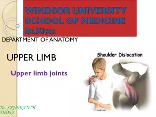
Dr. SREEKANTH THOTA. DEPARTMENT OF ANATOMY. UPPER LIMB. WINDSOR UNIVERSITY SCHOOL OF MEDICINE St.Kitts. SHOULDER . SHOULDER . The shoulder is the region of upper limb attachment to the trunk and neck. . Bones of the Shoulder Girdle .

E N D
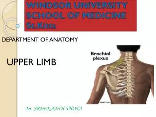
Dr. SREEKANTH THOTA DEPARTMENT OF ANATOMY UPPER LIMB WINDSOR UNIVERSITYSCHOOL OF MEDICINESt.Kitts SHOULDER
SHOULDER • The shoulder is the region of upper limb attachment to the trunk and neck.
Bones of the Shoulder Girdle • Clavicle and scapula, which form the pectoral girdle (shoulder girdle) • The proximal end of the humerus.
Muscles Shoulder Region
Anterior Axioappendicular Muscles of the Upper Limb • PECTORALIS MAJOR • PECOTORALIS MINOR • SUBCLAVIUS • SERRATUS ANTERIOR
PECTORALIS MAJOR • Origin: Clavicle, sternum, and upper six costal cartilages • Insertion : Lateral lip of bicipital groove of humerus • N. supply: Medial and lateral pectoral nerves • Action: Adducts arm and rotates it medially; clavicular fibers also flex arm
PECOTORALIS MINOR • Origin: Third, fourth, and fifth ribs • Insertion : Coracoid process of scapula • N. supply: Medial pectoral nerve • Action: Stabilizes scapula by drawing it inferiorly
SUBCLAVIUS • Origin: First costal cartilage • Insertion : Clavicle • N. supply: Nerve to subclavius • Action: Depresses the clavicle
SERRATUS ANTERIOR • Origin: Upper eight ribs • Insertion :Anterior surface of medial border of scapula • N. supply: Long thoracic nerve • Action: Protracts scapula and rotates scapula
Paralysis of the Serratus Anterior(winged scapula ) Due to loss of Innervation-long thoracic nerve • When the serratus anterior is paralyzed owing to injury to the long thoracic nerve, the medial border of the scapula moves laterally and posteriorly away from the thoracic wall, giving the scapula the appearance of a wing.
Posterior Axioappendicular Muscles • The posterior axioappendicular muscles attach the superior appendicular skeleton (of the upper limb) to the axial skeleton (in the trunk). • 1.Trapezius • 2.Latissimus dorsi • 3. Rhomboid major and minor • 4. Levator scapulae
Trapezius • Origin: Occipital bone, ligamentumnuchae, spine of seventh cervical vertebra, spines of all thoracic vertebrae • Insertion : Upper fibers into lateral third of clavicle; middle and lower fibers into acromion and spine of scapula • N. supply: Spinal part of accessory nerve (motor) • Action: Upper fibers elevate the scapula; middle fibers pull scapula medially; lower fibers pull medial border of scapula downward
Injury of the Accessory Nerve (CN XI) • The primary clinical manifestation of accessory nerve palsy is a marked ipsilateral weakness when the shoulders are elevated (shrugged) against resistance.
Latissimusdorsi • Origin: Iliac crest, lumbar fascia, spines of lower six thoracic vertebrae, lower three or four ribs, and inferior angle of scapula • Insertion: Floor of bicipital groove of humerus • N. supply:Thoracodorsal nerve
Latissimusdorsi Action: Extends, adducts, and medially rotates the arm
Injury of the Thoracodorsal Nerve • Surgery in the inferior part of the axilla puts the thoracodorsal nerve supplying the latissimusdorsi at risk of injury. • With paralysis of the latissimusdorsi, the person is unable to raise the trunk with the upper limbs, as occurs during climbing.
Levator Scapulae • Origin: Transverse processes of first four cervical vertebrae • Insertion: Medial border of scapula • N. supply:dorsal scapular nerve • Action:Elevates scapula
Rhomboids minor and major • N. supply: Dorsal scapular nerve • Action: Retracts scapula
Scapulohumeral Muscles • Scapulohumeral muscles: • The six scapulohumeral muscles are relatively short muscles that pass from the scapula to the humerus and act on the glenohumeral joint. • 1.Deltoid • 2.Teres major • 3.Teres minor • 4.Supraspinatus • 5.Infraspinatus • 6.Subscapularis
Deltoid • Origin: Lateral third of clavicle, acromion, spine of scapula • Insertion: Middle of lateral surface of shaft of humerus • N. supply: Axillary nerve
Deltoid Action: Anterior part: flexes and medially rotates armMiddle part: abducts armPosterior part: extends and laterally rotates arm
Testing deltoid muscle • The examiner resists the patient's abduction of the limb by the deltoid. • If the deltoid is acting normally, contraction of the middle part of the muscle can be palpated.
Injury to the Axillary Nerve The deltoid is a common site for the intramuscular injection of drugs. The axillary nerve runs transversely under cover of the deltoid at the level of the surgical neck of the humerus. The deltoid atrophies when the axillary nerve (C5 and C6) is severely damaged.
Teres Major • Origin: Lower third of lateral border of scapula • Insertion:Medial lip of bicipital groove of humerus • N. supply:Lowersubscapular nerve • Action:Adducts and medially rotates arm
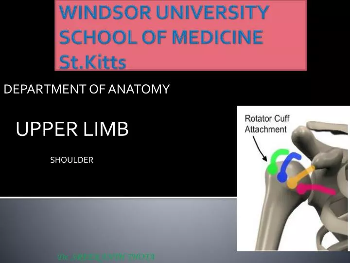
Rotator Cuff Muscles • Four of the scapulohumeral muscles supraspinatus, infraspinatus, teres minor, and subscapularis(referred to as the SITS muscles) are called rotator cuff muscles because they form a musculotendinous rotator cuff around the glenohumeral joint.
Teres minor • Origin: Middle part of lateral border of scapula • Insertion: Inferior facet of greater tubercle of humerus • N. supply:Axillary nerve • Action:Laterally rotates arm and stabilizes shoulder joint
Supraspinatus • Origin:Supraspinousfossa of scapula • Insertion: Superior facet of greater tubercle of humerus • N. supply:Suprascapular nerve • Action: Initiates and assists deltoid in abduction of arm and acts with rotator cuff muscles.
Infraspinatus • Origin:Infraspinousfossa of scapula • Insertion: Middle facet of greater tubercle of humerus • N. supply: Suprascapular nerve • Action: Laterally rotates arm and stabilizes shoulder joint
Subscapularis • Origin:Subscapularfossa (most of anterior surface of scapula) • Insertion: Lesser tubercle of humerus • N. supply: Upper and lower subscapular nerves • Action: Medially rotates and adduct arm
Rotator Cuff Injuries and the Supraspinatus • Trauma may tear or rupture one or more of the tendons of the SITS muscles; that of the supraspinatus is most commonly involved.
Triangle of Auscultation • Medial Border: • Trapezius muscle • Lateral Border: • scapula • Inferior Border (base): • LatissimusDorsi • It is an area of little muscle and hence a good place to listen to the posterior segment of the lungs
Arteries and nerves associated with gateways in the posterior scapular region
QUADRANGULAR SPACE: • Superior border: • Teres Minor • Inferior border: • Teres Major • Lateral border: • Surgical neck of the humerus • Medial border: • Long Head of Triceps
CONTENTS • 1- Axillary Nerve • 2- Posterir Circumflex Humeral Artery
Quadrilateral Space Syndrome • Quadrilateral space syndrome usually happens from overuse, especially with overhead sports like throwing and swimming. The syndrome can also be caused by an injury, like a shoulder dislocation.
TRIANGULAR [upper] space • LATERAL BORDER: • Long Head of the Triceps • Upper border: • Teres Minor and subscapularis • Lower border: • Teres Major • CONTENTS • Circumflex Scapular Branch • of the Subscapular artery
Lower Triangular Space • Medially • long head of the triceps brachii • Latteraly • shaft of the humerus • Superiorly • teres major
Contents • 1-Radial nerve • 2-Profunda brachii artery