Download
1 / 23
310 likes | 762 Views
Chapter 11: Nervous System Basics and Nervous System Tissues. Santiago Ramon Y. Cajal (1852-1934) Founding Scientist in the Modern Approach to Neuroscience. Received Nobel Prize in 1906. Figure 11.1: The nervous system’s functions, p. 388. Sensory input. Integration. Motor output.

E N D
Chapter 11: Nervous System Basics and Nervous System Tissues
Santiago Ramon Y. Cajal (1852-1934) Founding Scientist in the Modern Approach to Neuroscience. Received Nobel Prize in 1906
Figure 11.1: The nervous system’s functions, p. 388. Sensory input Integration Motor output
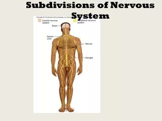
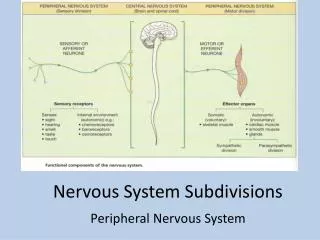
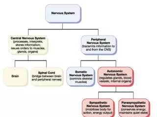
Figure 11.2: Levels of organization in the nervous system, p. 389. Key: Brain Central nervous system (CNS) Brain and spinal cord Integrative and control centers = Sensory (afferent) division of PNS = Motor (efferent) division of PNS Key: = Structure = Function Visceral sensory fiber Central nervous system (CNS) Peripheral nervous system (PNS) Cranial nerves and spinal nerves Communication lines between the CNS and the rest of the body Parasympathetic motor fiber of ANS Sympathetic motor fiber of ANS Visceral organ Spinal cord Skin Somatic sensory fiber Sensory (afferent) division Somatic and visceral sensory nerve fibers Conducts impulses from receptors to the CNS Motor (efferent) division Motor nerve fibers Conducts impulses from the CNS to effectors (muscles and glands) Motor fiber of somatic nervous system Skeletal muscle Sympathetic division Mobilizes body systems during activity Autonomic nervous system (ANS) Visceral motor (involuntary) Conducts impulses from the CNS to cardiac muscles, smooth muscles, and glands Somatic nervous System Somatic motor (voluntary) Conducts impulses from the CNS to skeletal muscles Peripheral nervous system (PNS) Parasympathetic division Conserves energy Promotes housekeeping functions during rest (b) (a)
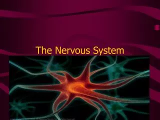
Figure 11.3: Neuroglia, p. 390. Capillary Neuron (b) Microglial cell (a) Astrocyte Nerve fibers Myelin sheath Fluid-filled cavity Process of oligodendrocyte Brain or spinal cord tissue (c) Ependymal cells Cell body of neuron (d) Oligodendrocyte Satellite cells Schwann cells (forming myelin sheath) Nerve fiber (e) Sensory neuron with Schwann cells and satellite cells
Figure 11.4: Structure of a motor neuron, p. 392. Cell body (biosynthetic center and receptive region) Dendrites (receptive regions) Neuron cell body Nucleus Dendritic spine (a) Axon (impulse generating and conducting region) Impulse direction Nucleolus Node of Ranvier Nissl bodies Axon terminals (secretory component) Axon hillock Schwann cell (one inter- node) Neurilemma (sheath of Schwann) Terminal branches (telodendria) (b)
Figure 11.5: Relationship of Schwann cells to axons in the PNS, p. 394. Schwann cell cytoplasm Schwann cell plasma membrane Axon Myelin sheath Schwann cell nucleus (a) Schwann cell cytoplasm Axon Neurilemma (b) (d) Neurilemma Myelin sheath (c)
Figure 11.6: Operation of gated channels, p. 398. Neurotransmitter chemical attached to receptor Receptor Na+ Na+ Chemical binds K+ K+ Closed Open (a) Chemically gated ion channel Na+ Na+ Membrane voltage changes Closed Open (b) Voltage-gated ion channel
Figure 11.7: Measuring membrane potential in neurons, p. 399. Voltmeter Plasma membrane Ground electrode outside cell Microelectrode inside cell Axon Neuron
Figure 11.8: The basis of the resting membrane potential, p. 399. Cell exterior Na+ Na+ 15 mM Cell interior Na+ Na+ K+ 150 mM ion Diffusion Na+–K+ pump us Diff Cl– 10 mM -70 mV Na+ Na+ A– 100 mM Na+ 150 mM K+ Na+ Plasma membrane A– 0.2 mM Na+ K+ K+ 5 mM Cl– 120 mM K+ Cell interior Cell exterior K+ K+
Figure 11.9: Depolarization and hyperpolarization of the membrane, p. 400. Depolarizing stimulus Hyperpolarizing stimulus +50 +50 Inside positive 0 0 Inside negative Depolarization Membrane potential (voltage, mV) Membrane potential (voltage, mV) –50 –50 Resting potential –70 –70 Resting potential Hyper- polarization –100 –100 0 1 2 3 4 5 6 7 0 1 2 3 4 5 6 7 Time (ms) Time (ms) (a) (b)
Figure 11.10: The mechanism of a graded potential, p. 401. Depolarized region Stimulus Plasma membrane (a) Depolarization (b) Spread of depolarization
Figure 11.11: Changes in membrane potential produced by a depolarizing graded potential, p. 402. Active area (site of initial depolarization) Membrane potential (mV) – 70 Resting potential Distance (a few mm)
Figure 11.12: Phases of the action potential and the role of voltage-gated ion channels, p. 403. Outside cell Outside cell Na+ Na+ Inside cell K+ Inside cell K+ Repolarizing phase: Na+ channels inactivating, K+ channels open Depolarizing phase: Na+ channels open 2 Action potential +30 3 0 Relative membrane permeability 2 PNa Membrane potential (mV) PK Threshold –55 1 1 4 –70 0 1 2 3 4 Time (ms) Outside cell Sodium channel Potassium channel Outside cell Na+ Na+ Activation gates K+ Inside cell K+ Inside cell Inactivation gate Hyperpolarization: K+ channels remain open; Na+ channels resetting 4 1 Resting state: All gated Na+ and K+ channels closed (Na+ activation gates closed; inactivation gates open)
Figure 11.13: Propagation of an action potential (AP), p. 405. Voltage at 2 ms +30 Membrane potential (mV)) Voltage at 0 ms Voltage at 4 ms –70 (a) Time = 0 ms (b) Time = 2 ms (c) Time = 4 ms Resting potential Peak of action potential Hyperpolarization
Figure 11.14: Relationship between stimulus strength and action potential frequency, p. 406. Action potentials +30 Membrane potential (mV) – 70 Stimulus amplitude Threshold Voltage 0 Time (ms)
Figure 11.15: Refractory periods in an AP, p. 406. Absolute refractory period Relative refractory period Depolarization (Na+ enters) +30 0 Repolarization (K+ leaves) Membrane potential (mV) After-hyperpolarization –70 Stimulus 0 1 2 3 4 5 Time (ms)
Figure 11.16: Saltatory conduction in a myelinated axon, p. 407. Node of Ranvier Cell body Myelin sheath Distal axon
Figure 11.17: Synapses, p. 409. Cell body Dendrites Axodendritic synapses Axosomatic synapses Axoaxonic synapses Axon (a) Axon Axosomatic synapses Soma of postsynaptic neuron (b)
Figure 11.18: Events at a chemical synapse in response to depolarization, p. 410. Neurotransmitter Na+ Ca2+ Axon terminal of presynaptic neuron Receptor Action Potential 1 Postsynaptic membrane Mitochondrion Postsynaptic membrane Axon of presynaptic neuron Ion channel open Synaptic vesicles containing neurotransmitter molecules 5 Degraded neurotransmitter Na+ 2 Synaptic cleft 3 4 Ion channel closed Ion channel (closed) Ion channel (open)
Figure 11.19: Postsynaptic potentials, p. 412. +30 +30 0 0 Threshold Membrane potential (mV) Threshold Membrane potential (mV) –55 –55 –70 –70 10 20 Time (ms) 10 20 Time (ms) (b) Inhibitory postsynaptic potential (IPSP) (a) Excitatory postsynaptic potential (EPSP)
Figure 11.24: Types of circuits in neuronal pools, p. 422. Input Input Input Input 1 Input 2 Input 3 Output Output Output Output (a) Divergence in same pathway (b) Divergence to multiple pathways (c) Convergence, multiple sources (d) Convergence, single source Input Input Output Output (e) Reverberating circuit (f) Parallel after-discharge circuit
Figure 11.25: A simple reflex arc, p. 423. Integration center Sensory neuron Stimulus Receptor Interneuron Response Effector Motor neuron Spinal cord (CNS)