Download
1 / 25
250 likes | 451 Views
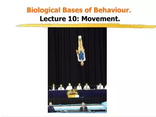
Biological Bases of Behaviour. Lecture 10: Movement. Learning Outcomes. By the end of this lecture you should be able to: 1 . Outline the key anatomical components of the movement system. 2 . Describe the effects of damage to each of the components.

E N D
Learning Outcomes. • By the end of this lecture you should be able to: • 1. Outline the key anatomical components of the movement system. • 2. Describe the effects of damage to each of the components. • 3. Explain how the various components of the system are organised.
Anatomy of Movement. • The motor system can be divided into a number of subsystems: • 1. Muscles • 2. Spinal cord • 3. Cerebellum • 4. Reticular formation • 5. Basal ganglia • 6. Cortex • They share many interconnections with each other. • Remember that each hemisphere of the brain controls the trunk of the body on the same side, and the arms and legs of the opposite side.
1. Muscles. • A part of the body that can move is called an effector. • A distal effector is far from the body centre (hands, feet). • A proximal effector is centrally located (waist, neck, eyes). • Muscles are attached to the skeleton at joints and can only move in one direction. • In order to make an arm extend and flex, it requires a pair of muscles working in opposition enabling the effector to flex and extend. • E.g: activating the biceps flexes the forearm, activating the triceps extends the forearm.
Muscle Action. Tricep relaxes Bicep contracts Bicep relaxes Tricep contracts Kalat (2001) p225
Muscles (continued). • A neuromuscular junction is a synapse where a motor neuron synapses with a muscle fibre, all such connections are excitatory and release acetylcholine. • The muscles interact with the nervous system via the alpha motor neurons which originate in the ventral section of the spinal cord. • In vertebrates there are three types of muscles: • Smooth muscles: control movements of internal organs. • Skeletal muscles: control the movement of the body parts. • Cardiac muscles: control the heart.
Muscle Control. • Muscles are controlled by proprioceptors which are specialised receptors sensitive to the position and movement of the body. • They detect the stretch and tension of a muscle and send messages to the spinal cord to enable it to adjust its signals to the muscles. There are two main types: • a) Muscle spindle: a stretch receptor lying parallel to the muscle. When stretched it sends a message to a motor neuron in the spinal cord which in turn relays a message to the muscle causing a contraction. E.g the knee-jerk reflex. • B) Golgi tendon organ: located in the tendons at both ends of the muscle. It acts as a brake against excessive contractions by inhibiting the motor neurons in the spinal cord.
Proprioceptors Spinal cord Motor neurons Sensory neurons Muscle Muscle spindle Golgi tendon organ Kalat (2001) p227
Disorders of the Motor Neurons. • a) Myasthenia Gravis: The immune system progressively attacks acetylcholine at the neuromuscular junction. • This leads to progressive muscle weakness and rapid fatigue apparent after short periods of exercise. • Drugs such as Physostigmine (an acetylcholine agonist) alleviate the symptoms. • b) Multiple Sclerosis: A common diseases characterised by the loss of myelin surrounding sensory and motor neurons. • The myelin loss is patchy but a sclerotic plaque forms which severely impairs the functioning of the neuron. • Onset is rapid with first symptoms being limb weakness, disorders of vision, tremors, vertigo. • There is no treatment as yet.
2.Spinal Cord. • This cord distributes motor fibres to the effectors and collects sensory information to be passed to the brain. • It is protected by 24vertebrae. • Small bundles of fibres emerge from the spinal cord, groups of these bundles fuse to form the dorsal and ventral roots which form the spinal nerves. Dorsal roots Ventral roots
Effects of Damage to the Spinal Cord. • Dorsal root damage: normal movement is possible but sensory information is lost. • Ventral root damage: sensory information is received but movement is lost. • Paraplegia: Lower limbs are paralysed. • Hemiplegia: One side of the body is paralysed. • Quadraplegia: All four limbs and the trunk of the body are paralysed - usually caused by a broken neck.
3. Cerebellum. • This structure contains more neurons than the rest of the brain. • It receives input from the spinal cord, the sensory systems, and from the cortex. • It co-ordinates muscle activity, maintains balance, and plays a role in motor skill learning.
Effects of Cerebellum Damage. • Cerebral palsy: Caused by lack of oxygen during the birth process. • Movements becomes jerky, erratic, and uncoordinated, and movement sequencing becomes problematic. • Also affected is speech, control of eye movements, writing and even simple alternating movements (such as clapping) become difficult. • Alcohol intoxication: It is one of the first areas of the brain to show the effects of alcohol intoxication. • Symptoms are slurred speech, clumsy motor control, loss of balance, and inaccurate eye movements.
4. Basal Ganglia. • This comprises a set of interconnected nuclei in the forebrain which includes: • Caudate nucleus. • Globus pallidus. • Substantia nigra. • Subthalamic nucleus. • Putamen. • The caudate nucleus and putamen receive sensory input from the thalamus and cortex, while the globus pallidus sends information to the primary motor cortex via the thalamus.
Basal Ganglia. amygdala Substantia nigra
Effects of Damage to the Basal Ganglia • The basal ganglia have rich connections to the cerebral cortex and subcortical nuclei. • They not only contribute to movement but they also influence cognitive functioning (though exactly how is not yet known). • Damage to them produces a variety of changes in movement though two main problems emerge: • Akinesia (an absence of spontaneous movement) as seen in Parkinson’s disease. • Hyperkinesia (rapid involuntary movements) as seen in Huntington’s disease.
5. Reticular Formation. • This consists of a large number of nuclei located in the core of the medulla, pons and midbrain which principally control muscle tone and posture. • Nuclei in the pons and medulla also control automatic movements such as vomiting, coughing and sneezing. • Other motor functions of this structure are still being discovered but it also seems to play a role in locomotion though it does not have a direct connection to the spinal cord.
6. Cerebral Cortex. • The role of certain regions of the cerebral cortex in motor activity was discovered in 1870 by Fritsch & Hitzig. • They electrically stimulated the exposed cortex of a dog and observed the co-ordinated movements of several muscles. • The cerebral cortex does not connect directly to the muscles, but sends axons to the medulla and spinal cord, which in turn send axons to the muscles. • So, unlike the spinal cord, the cortex is responsible for the overall planning of movements and not individual muscle contractions. • It is not responsible for automatic and involuntary movements e.g coughing, laughing, sneezing and gagging.
Cortical Motor Regions • There are several cortical regions controlling various aspects of movement: • Primary motor cortex: The main movement processing region, within which separate areas control different parts of the body. • Premotor cortex: Active during the preparations before a movement has begun (motor planning). • Supplementary motor area: Active during the preparation before a rapid series of voluntary movements. • Prefrontal cortex: Responds mostly to sensory signals that lead to movement. • Somatosensory cortex: Primary receiving area for touch and and is closely connected with the motor processing regions and spinal cord.
Cerebral Cortex (continued). Primary motor cortex Primary somatosensory cortex Central sulcus Supplementary motor cortex Premotor cortex Prefrontal cortex Kalat (2001) p232
Effects of Damage to the Cerebral Cortex. • By investigating patients with various types of brain damage we can see how the various components of motor performance may be affected. Examples: • Lesions to primary motor cortex (ie from a stroke) result in loss of voluntary movements on the contralateral (opposite) side of the body. • Apraxia is the specific loss of the ability to plan and correctly perform co-ordinated motor skills, mainly as a result of damage to the supplementary motor area. • Patients can move muscles, and walk on command but can no longer link gestures to a coherent act, or to recognise the appropriate use of an object even though they can recognise what an object is.
Cortical Control of Movement. • The cortex can regulate the activity of spinal neurons in direct and indirect ways: • a) Pyramidal system: Consists mainly of axons from primary motor cortex and adjacent areas. • These axons descend to the medulla where they decussate (cross over) in distinctive swellings called pyramids. • The fibres then split as they enter the spinal cord, forming the: • Dorsolateral tract: controls peripheral body movements (fingers, toes etc). • Ventromedial tract: controls movements at the midline (back and neck etc). • This system is also referred to as the corticospinal tract.
Cortical Control of Movement (continued). • b) Extrapyramidal system: This consists of all the movement-controlling areas other than the pyramidal system. • Axons from the basal ganglia and diffuse areas of the cortex converge onto the red nucleus, the reticular formation, and the vestibular nucleus. • Each of these areas then sends axons to the medulla and spinal cord.
Hierarchy of Movement Control. • Most movements rely on both pyramidal and extrapyramidal systems, and on dorsomedial and ventrolateral tracts. There is a hierarchy of motor control: • Lowest level: The spinal cord which provides a point of contact between the nervous system and the muscles, and also controls reflexive movements. • Middle level: The motor cortex and brainstem structures (plus the the cerebellum and basal ganglia) translate a specific set of action goals into movement via their communication with the spinal cord. • Highest level: Premotor and cortical association areas which are concerned with the planning and organisation of movement based on current perceptual information and previous experience.
Bibliography. • Carlson, N.R. (1994). Physiology of Behaviour. • Gazzaniga, M.S., Ivry, R.B., & Mangun, G.R. (1998). Cognitive Neuroscience. • Kalat, J.W. (1995). Biological Psychology.