Download
1 / 40
430 likes | 632 Views
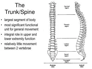
Muscles of the Trunk and Limbs. Muscles of the Trunk. The trunk muscles are involved in: forming the “girdle” of the abdominal body wall moving the vertebral column (extension and flexion) moving the ribs, head, and arms. Anterior Muscles. Pectoralis Major

E N D
Muscles of the Trunk The trunk muscles are involved in: • forming the “girdle” of the abdominal body wall • moving the vertebral column (extension and flexion) • moving the ribs, head, and arms
Anterior Muscles Pectoralis Major • A large fan-shaped muscle covering the upper part of the chest. • Originates at the clavicle and sternum. • Attaches at the proximal end of the humerus. • Functions to adduct and flex the arm.
Intercostal Muscles • Deep muscles found between the ribs. • There are 2 types: 1.External intercostals which help raise the ribs for breathing in air. 2.Internal intercostals which depress the rib cage and helps move air out of the lungs when you exhale forcibly.
Muscles of the Abdominal Girdle • Are made up of: • Rectus abdominus • External obliques • Internal obliques • Transverse abdominus • Form a natural “girdle” that holds in your guts. • The muscles run in different directions forming an especially strong muscular wall.
Transverse Abdominus External Oblique Internal Oblique Rectus Abdominus
Rectus Abdominus • The most superficial muscle set. • Runs in pairs from the pubis to the lower ribs. • The main function is to flex the vertebral column. • They also: • compress the abdomen during defecation • compress the abdomen during childbirth
External Obliques • Paired superficial muscles that make up the lateral walls of the abdomen. • Their fibers run downward and medially from the lower ribs and insert into the iliac crest. • They function to: • flex the vertebral column • rotate the trunk
Internal Obliques • Paired muscles found deep to the external obliques. • The fibers run downward and lateral from the ribs to the iliac crest. • Their function is the same as those of the external obliques. Internal Obliques
Transverse Abdominis • The deepest muscle of the abdomen. • Has fibers that run horizontally across the abdomen. • This muscle compresses the abdominal contents. Transverse abdominus
Posterior Muscles Trapezius • The most superficial muscles of the posterior neck and upper trunk. • The left and right side together form a diamond or kite-shaped muscle mass. • They originate from the occipital bone and the vertebrae and they run laterally to the scapula and clavicle. • They elevate, depress, adduct and generally move the scapula.
Latissimus Dorsi • The large, flat muscle pair that covers the lower back. • It stretches from the lower spine and ilium and inserts into the proximal end of the humerus. • It functions to extend, hyper-extend, and adduct the humerus.
Erector Spinae • The paired deep muscles found parallel to either side of the vertebral column. • Function as back extensors. Erector Spinae
Deltoid • Fleshy, triangle-shaped muscles that form the rounded shape of the shoulders. • They run from the shoulder girdle to the proximal humerus • The primary function is arm abduction.
STOP HERE Muscles of Limbs are next.
Muscles of the Upper Limbs These muscles fall into 3 groups: • The muscles that move across the shoulder and insert into the humerus (we’ve already mentioned these). 2. The muscles that cause movement at the elbow joint. • The muscles that cause flexion and extension of the wrist and fingers
Biceps Brachii The best known arm muscle, it bulges when the elbow is flexed. • Has 2 parts that originate at the shoulder girdle and it inserts at the proximal radius. • The primary function is flexion, but it also supinates the forearm.
Brachialis • This muscle lies deep to the biceps muscle. • It aides in elbow flexion. Brachioradialis • A weak muscle that arises from the humerus and inserts into the distal radius. • Flexes semi-pronated and semi-supinated forearm.
Triceps Brachii The only muscle on the posterior portion of the humerus. • It has 3 parts that arise from the shoulder girdle and the proximal humerus and it inserts into the olecranon process of the ulna. • The function is to extend the forearm by being the antagonist of the biceps brachii.
Muscles of Lower Limbs These muscles act on the lower limb and cause movement at: • Hip • Knee • Foot joints Are specialized for walking and balance
Muscles that Move the Hip Gluteus Maximus • A superficial muscle of the hip • Forms the fleshy part of the buttock • Originate at sacrum and ilium inserts into femur • Functions as an extensor to bring the thigh in a straight line with the pelvis • Not very important for walking, more important for jumping, standing up, and climbing hills or stairs.
Gluteus Medius • Runs from the iliac crest to the femur deep to the gluteus maximus. • Functions as a hip abductor and is important in steadying the pelvis during walking.
Adductor Muscles • These muscles form the mass at the medial side of the each thigh. • They originate on the pelvis and insert on the proximal femur. • Their main function is to adduct or press the thighs together. • Because they do not work against gravity, they are likely to become flabby easily.
Muscles that Move the Knee Hamstring Group • A group of 3 muscles that form the mass of the posterior thigh. • Consists of the: 1. Biceps femoris 2. Semimembranosus 3. Semitendinosus • They originate on theischiumand insert at the proximal tibia. Main function is to flex the knee
Sartorius • The most superficial muscle of the thigh. • It runs obliquely across the thigh from the anterior iliac crest to the medial side of the tibia. • The primary function is to cross your legs.
Quadriceps Group • A group of muscles that makes up the muscle mass on the anterior thigh • Consists of 4 muscles: • Rectus femoris • Vastus lateralis • Vastus medialis • Vastus intermedius • The rectus femoris originates at the pelvis and the vastus muscles originate at the proximal femur. All muscles insert into the tibial tuberosity. • Functions to extend the knee and to flex the hip.
Rectus Femoris Vastus Lateralis Vastus Intermedius Vastus Medialis
Muscles that Move the Foot Tibialis Anterior • A superficial muscle on the anterior leg. • Runs parallel to the anterior crest from the proximal tibia and inserts via tendons to the tarsal bones. • Functions to dorsiflex and invert the foot and move toe #1
Extensor Digitorum Longus • A smaller muscle that runs lateral to the tibialis anterior. • It arises from the lateral tibial condyle and it inserts into phalanges of toes 2-5. • It functions to extend toes 2-5 and dorsiflex the foot.
Fibularis Muscles • A group of muscles found on the lateral part of the leg. • Consists of: • Fibularis Longus • Fibularis Brevis • Fibularis Tertius • The function is plantar flexion and eversion of the foot.
Gastrocnemius • A 2 – bellied muscle that forms the curved calf of the posterior leg. • Each of the heads arises from each side of the distal femur and they join and insert by the calcaneal (Achilles) tendon into the heel of the foot. • The main function is plantar flexion.
Soleus • A fleshy muscle that lies deep to the gastrocnemius. • It arises on the proximal tibia and inserts at the heel. • It aides in plantar flexion.