Microbiology
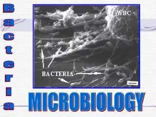
Microbiology. Review of Gram Stain Selective and differential Media. Gram Positive Bacteria. Staphylococcus aureus Streptococcus salivarius Bacillus subtilis Staphylococcus epidermidis. Gram Positive Cell Wall. Gram Negative Bacteria. E. coli Pseudomonas aeruginosa Proteus vulgaris

Microbiology
E N D
Presentation Transcript
Microbiology Review of Gram Stain Selective and differential Media
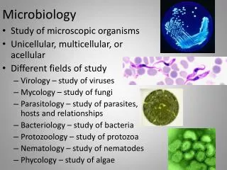
Gram Positive Bacteria • Staphylococcus aureus • Streptococcus salivarius • Bacillus subtilis • Staphylococcus epidermidis
Gram Negative Bacteria • E. coli • Pseudomonas aeruginosa • Proteus vulgaris • Enterobacter aerogenes
The Smear • 1. Label the frosted side of your slide with your initials, the name of the organism, and the date. On this side draw a circle in the clear section of the slide. • 2. Turn the slide over. You will make your smear on this side. *****If you are using broth follow these directions • Flame your inoculating loop. • Use aseptic technique and remove the top of the culture tube, flame the mouth or the culture tube, and dip the loop into the broth. Make sure that the loop is filled. • Transfer the loopful of broth and bacteria to the slide. Using a circular motion, spread the broth on the slide. • This is now a " smear" • Allow the smear to dry • When the smear has been allowed to " air dry" , pass the smear through the flame to " heat fix" - Heat fixation causes the proteins and cell parts to coagulate and stick to the slide. • Let the slide cool.
Simple Stain • Simple Stains- Crystal violet and methylene blue • Place a drop(s) of stain over the smear. Make sure it covers the entire area of the smear. Leave the stain on the smear for one minute. Rinse with water from the bottle. • Refer to page 64 for cellular morphology
Gram Stain • Gram Stain- See Gram Stain Directions on separate page. Please refer to pages 71-73 in your laboratory manual. • All staining work is to be done at the sink • Care should be taken to work directly over the sink • Place drop(s) of crystal violet stain on the smear ( 1 minute) • Rock or roll the slide to cover the area • Use the water bottle to drip water down the slide • Place drop(s) of iodine on the slide ( 1 minute) • Place drops of alcohol on the slide 10 seconds ( KEY – do not leave on longer than 10 seconds or it will decolorize) • Place drop(s) of saffranin on the slide for 1 minute • Rinse with water from the bottle • Let the slide air dry
Gram Staininig examples • http://www.uphs.upenn.edu/bugdrug/antibiotic_manual/Gram3.htm • http://www.uphs.upenn.edu/bugdrug/antibiotic_manual/Gramstains/small/tocframeset1.htm
Reagents • Crystal Violet (the Primary Stain) • Iodine Solution (the Mordant) • Decolorizer (ethanol is a good choice) • Safranin (the Counterstain) • Water (preferably in a squirt bottle)
Selective Media • Selective media is used to isolate specific groups of bacteria • They include ingredients that inhibit the growth of one type of bacterium and permit the growth of another
Differential Media • Specialized media assists in the identification of bacteria • Media contains ingredients that are essential for the completion of specific biochemical reactions • Assist in the identification of bacteria that are closely related by observing both the bacterial growth and the appearance of the agar
Mannitol Salt Agar • This media contains • 7.5% salt which is inhibitory to many organisms( organisms that can exist in a high salt environment are described as halophilic) Staphlococci are uniue in this respect • It also contains a carbohydrate mannitol – which is a sugar that staphylococci are capable of utilizing for energy through the process of fermentation
Phenol Red • Phenol red is a pH indicator for detecting the acid that is produced by fermentation of mannitol • The phenol red changes to yellow in the presence of acid. • The staphylococci exhibit this characteristic color
MacConkey • Crystal violet is present in this agar • It inhibits the growth of Gram Positive Bacteria • It does permits the growth of Gram Negative Bacteria
MacConkey( continued) • It contains the sugar lactose • Bile salts • The pH indicator neutral red
MacConkey as Differential • This agar distinguishes between enteric bacteria on the basis of their ability to ferment lactose sugar. • If a bacterium is capable of fermenting lactose it produces pinkish colonies. Non fermenter colonies are purple
Eosin – Methylene Blue( EMB) • Lactose sugar • Dyes eosin and methylene blue permit differentiation between enteric lactose fermenters and non fermenters • It is also possible to identify E. coli Because it produces a characteristic blue-black color with a metallic green sheen
PEA( Phenylethyl alchohol agar) • This media is used to identify gram positive bacteria • The phenyl ethyl alchohol inhibits the growth of gram negative organisms due to the construction of their cell wall. • If gram negative organisms grow their colonies are smaller than on other media