Download
1 / 128
1.29k likes | 1.33k Views
Dr. Michael P. Gillespie explains the essential components and features of the human axial skeleton, focusing on the cranium, temporal bones, occipital bone, vertebrae, ribs, and sternum. Learn about the structure and function of these key skeletal elements.

E N D
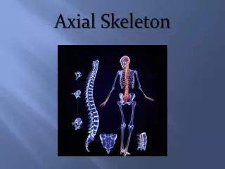
AXIAL SKELETONOSTEOLOGY AND ARTHROLOGY Dr. Michael P. Gillespie
HUMAN SKELETON: ANTERIOR VIEW Dr. Michael P. Gillespie
HUMAN SKELETON: POSTERIOR VIEW Dr. Michael P. Gillespie
RELATIVE LOCATION OR REGION WITHIN THE AXIAL SKELETON Dr. Michael P. Gillespie The definitions assume a person is in the anatomic position.
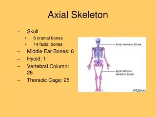
COMPONENTS OF THE AXIAL SKELETON • Cranium • Vertebrae • Ribs • Sternum Dr. Michael P. Gillespie
CRANIUM • The cranium encases and protects the brain. • It houses several sensory organs. • Eyes, ears, nose and vestibular system. • Only the temporal and occipital bones are relevant to our study of kinesiology. Dr. Michael P. Gillespie
OSTEOLOGIC FEATURES OF THE CRANIUM • Temporal Bone • Mastoid process • Occipital Bone • External occipital protruberance • Superior nuchal line • Inferior nuchal line • Foramen magnum • Occipital condyles • Basilar part Dr. Michael P. Gillespie
TEMPORAL BONES • The two temporal bones form part of the lateral external surface of the skull immediately surrounding and including the external acoustic meatus. • The mastoid process is just posterior to the ear and serves as an attachment point to many muscles (i.e. sternocleidomastoid and longissimus). Dr. Michael P. Gillespie
OCCIPITAL BONE • The occipital bone forms the posterior base of the skull. • The external occipital protruberance (EOP) is a palpable midline point. It is an attachment point for the ligamentum nuchae and the medial part of the upper trapezius muscle. • The superior nuchal line extends laterally from the EOP to the base of the mastoid process of the temporal bone. This line serves as the attachment point for several muscles of the neck (i.e. trapezius and splenius capitis). • The inferior nuchal line marks the anterior edge of the attachment of the semispinalis muscle capitis muscle. • The foramen magnum is a large circular hole at the base of the occipital bone. It serves as a passageway for the spinal cord. • Occipital condyles project from the anterior-lateral margins of the foramen magnum forming the convex component of the atlanto-occipital joint. • The basilar part of the occipital bone lies just anterior to the anterior rim of the foramen magnum. Dr. Michael P. Gillespie
LATERAL VIEW OF THE SKULL Dr. Michael P. Gillespie
INFERIOR VIEW OF THE OCCIPITAL AND TEMPORAL BONES Dr. Michael P. Gillespie
VERTEBRAE • The vertebrae provide stability throughout the trunk and neck. They protect the spinal cord, ventral and dorsal roots, and exiting spinal nerve roots. • 3 sections of the vertebra • Vertebral body (anterior) • Transverse and spinous processes (posterior) – posterior elements (neural arch, vertebral arch) • Pedicles – bridges that connect the body with the posterior elements – transfer muscle forces applied to the posterior elements forward across the vertebral body and intervertebral discs. Dr. Michael P. Gillespie
MAJOR PARTS OF A MIDTHORACIC VERTEBRA • Table 9-2 • Major parts of a Midthoracic Vertebra • Chapter 9 Page 311 Dr. Michael P. Gillespie
ESSENTIAL CHARACTERISTICS OF A VERTEBRA Dr. Michael P. Gillespie
ESSENTIAL CHARACTERISTICS OF A VERTEBRA Dr. Michael P. Gillespie
RIBS • Twelve pairs of ribs enclose the thoracic cavity forming a protective cage for the cardiopulmonary organs. • The rib head and tubercle articulate with the thoracic vertebrae forming two synovial joints: • Costocorporeal (costovertebral) • Costotransverse • These joints anchor the posterior end of a rib to its corresponding vertebra. • The anterior end of a rib consists of flattened hyaline cartilage. Dr. Michael P. Gillespie
TYPICAL RIB Dr. Michael P. Gillespie
STERNUM • Three parts • Manubrium (Latin – handle) • Body • Xiphoid process (Greek – sword) • The manubrium fuses with the body of the sternum at the manubriosternal joint (a cartilaginous joint that often ossifies later in life). • The xiphoid process is connected to the sternum by fibrocartilage at the xiphisternal joint that often fuses by 40 years of age. • Sternoclavicular joints. • Sternocostal joints. Dr. Michael P. Gillespie
OSTEOLOGIC FEATURES OF THE STERNUM • Osteologic Features of the Sternum • Manubrium • Jugular notch • Clavicular facets for sternoclavicular joints • Body • Costal facets for sternocostal joints • Xiphoid process • Intrasternal Joints • Manubriosternal joint • Xiphosternal joint Dr. Michael P. Gillespie
STERNUM Dr. Michael P. Gillespie
VERTEBRAL COLUMN • 33 vertebral bony segments divided into five regions. • Cervical • Thoracic • Lumbar • Sacral • Coccygeal Dr. Michael P. Gillespie
CURVATURES WITHIN THE VERTEBRAL COLUMN • When viewed from the side, the vertebral column shows four slight bends called normal curves. • Relative to the anterior aspect of the body, the cervical and lumbar curves are convex (bulging out), whereas the thoracic and sacral curves are concave (cupping in). • The curves in the vertebral column increases its strength, help maintain balance in the upright position, absorb shocks during walking, and help to protect the vertebrae from fracture. • Various conditions may exaggerate the normal curves of the vertebral column, or the column may acquire a lateral bend, resulting in abnormal curves. • The abnormal curves are kyphosis, lordosis, and scoliosis. Dr. Michael P. Gillespie
VERTEBRAL COLUMN Dr. Michael P. Gillespie
INCORRECT LABELING OF THE NORMAL CURVES Dr. Michael P. Gillespie
EXTENSION AND FLEXION OF THE VERTEBRAL COLUMN Dr. Michael P. Gillespie
LINE OF GRAVITY • The line of gravity acting on a person with ideal posture passes near the mastoid process of the temporal bone, anterior to the second sacral vertebra, just posterior to the hip, and anterior to the knee and ankle. • In the vertebral column, the line of gravity typically falls just to the concave side of the apex of each region’s curvature. • Ideal posture allows gravity to produce a torque that helps maintain the optimal shape of the spinal curvatures. • The external torque attributed to gravity is the greatest at the apex of each region: C4 and C5, T6, and L3. Dr. Michael P. Gillespie
LINE OF GRAVITY Dr. Michael P. Gillespie
COMMON POSTURAL DEVIATIONS Dr. Michael P. Gillespie
LIGAMENTOUS SUPPORT OF THE VERTEBRAL COLUMN • The vertebral column has extensive ligament support. • These ligaments limit motion, help maintain natural spinal curvatures, stabilize the spine, and protect the spinal cord and nerve roots. Dr. Michael P. Gillespie
LIGAMENTS: LATERAL VIEW Dr. Michael P. Gillespie
LIGAMENTS: ANTERIOR VIEW Dr. Michael P. Gillespie
LIGAMENTS: POSTERIOR VIEW Dr. Michael P. Gillespie
MAJOR LIGAMENTS OF THE VERTEBRAL COLUMN Dr. Michael P. Gillespie
MAJOR LIGAMENTS OF THE VERTEBRAL COLUMN Dr. Michael P. Gillespie
STRESS STRAIN CURVE LIGAMENTUM FLAVUM Dr. Michael P. Gillespie
PROMINENT LIGAMENTUM FLAVUM Dr. Michael P. Gillespie
CERVICAL REGION • Smallest and most mobile of the vertebrae, which facilitates the large range of motion of the head. • Transverse foramina are located in the transverse processes of the cervical spine through which the vertebral artery travels. Dr. Michael P. Gillespie
CERVICAL VERTEBRA: SUPERIOR VIEW Dr. Michael P. Gillespie
CERVICAL VERTEBRA: ANTERIOR VIEW Dr. Michael P. Gillespie
TYPICAL CERVICAL VERTEBRAE (C3 TO C6) • Small rectangular bodies. • The superior surfaces are concave side to side, with raised lateral hooks called uncinate processes (uncus means “hook”). • These form the uncovertebral joints (a.k.a. “joints of Luschka”). • Osteophytes can form around the margins of these joints which can reduce the size of the intervertebral foramen (IVF) and impinge upon exiting nerve roots. • Superior articular facets face posterior and superior, whereas the inferior articular facets face anterior and inferior. Dr. Michael P. Gillespie
CERVICAL VERTEBRA: POSTERIOR-LATERAL VIEW Dr. Michael P. Gillespie
CERVICAL VERTEBRAL COLUMN: LATERAL VIEW Dr. Michael P. Gillespie
ATYPICAL CERVICAL VERTEBRAE (C1, C2, & C7) • Atlas (C1) • Axis (C2) • “Vertebra Prominens” (C7) Dr. Michael P. Gillespie
ATLAS (C1) • The primary function is to support the head. • The atlas has large, palpable transverse processes, usually the most prominent of the cervical vertebrae. • The transverse processes serve as attachment points for muscles that move the cranium. Dr. Michael P. Gillespie
ATLAS Dr. Michael P. Gillespie
ATLAS (C1) Dr. Michael P. Gillespie
AXIS (C2) • The axis has an upwardly projecting dens (odontoid process) which provides a vertical axis of rotation for the atlas and head. Dr. Michael P. Gillespie
AXIS (C2) Dr. Michael P. Gillespie
AXIS (C2) Dr. Michael P. Gillespie