Understanding Neurons, Nervous Systems, and Brain Activity
Dive into the world of neurons, nervous systems, and brain activity. Explore how neurons fire signals, the anatomy of a neuron, nerve impulses, synapses, neurotransmitters, neural regulators, and neuroplasticity. Discover the complex relationship between the mind and the brain through PET scans. Unravel the mysteries of neural networks and the central and peripheral nervous systems.

Understanding Neurons, Nervous Systems, and Brain Activity
E N D
Presentation Transcript
Weekly Reading Guarantee • What is the action potential? In other words, do neurons fire half way? As in communicate half a signal? Or is it an all or nothing event? • Is there more than one nervous system? Are we conscious of everything our nervous system is doing?
Grounding this information in something coherentPhineas Gage http://www.youtube.com/watch?v=kc213mMSsjY
Neuron’s Parts • Soma: Cell body; body of the neuron. Receives messages and sends messages down axon • Axon: Fiber that carries information away from the cell body of a neuron • Axon terminals: Branches that link the dendrites and somas of other neurons • Dendrites: Receive messages from other neurons
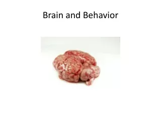
A neuron, or nerve cell. In the right foreground you can see a nerve cell fiber in cross section. The upper left photo gives a more realistic picture of the shape of neurons. Nerve impulses usually travel from the dendrites and soma to the branching ends of the axon. The nerve cell shown here is a motor neuron. The axons of motor neurons stretch from the brain and spinal cord to muscles or glands of the body. Fig. 2-1, p. 49
The interior of an axon. The right end of the top axon is at rest. Thus, it has a negative charge inside. An action potential begins when ion channels open and sodium ions (Na+) rush into the axon. In this drawing, the action potential would travel from left to right along the axon. In the lower axon, the action potential has moved to the right. After it passes, potassium ions (K+) flow out of the axon. This quickly renews the negative charge inside the axon so that it can fire again. Sodium ions that enter the axon during an action potential are pumped out more slowly. Removing them restores the original resting potential. Fig. 2-4, p. 51
The Nerve Impulse • Resting potential: Electrical charge of an inactive neuron • Threshold: Trigger point for a neuron’s firing • Action Potential: Nerve impulse: This is an all or nothing event, they cannot fire half-way
Electrical probes placed inside and outside an axon measure its activity. (The scale is exaggerated here. Such measurements require ultra-small electrodes, as described in this chapter.) The inside of an axon at rest is about –60 to –70 millivolts, compared with the outside. Electrochemical changes in a neuron generate an action potential. When sodium ions (Na+) that have a positive charge rush into the cell, its interior briefly becomes positive. This is the action potential. After the action potential, positive potassium ions (K+) flow out of the axon and restore its negative charge. Fig. 2-2, p. 50
More on Nerves • Ion channels: Tiny openings through the axon membrane • Negative after-potential: A drop in electrical charge below the resting potential • Synapse: Microscopic space between two neurons over which messages pass
A highly magnified view of a synapse. Neurotransmitters are stored in tiny sacs called synaptic vesicles (VES-ih-kels). When a nerve impulse reaches the end of an axon, the vesicles move to the surface and release neurotransmitters. These molecules cross the synaptic gap to affect the next neuron. The size of the gap is exaggerated here; it is actually only about one millionth of an inch. Some transmitter molecules excite the next neuron and some inhibit its activity. Fig. 2-5, p. 51
Neurotransmitters • Chemicals that alter activity in neurons; brain chemicals • Receptor site: Area on the surface of neurons and other cells that is sensitive to neurotransmitters or hormones
Neural Regulators • Neuropeptides: Regulate activity of other neurons • Enkephalins: Relieve pain and stress; similar to endorphins • Endorphins: Released by pituitary gland; also help to relieve pain • Placebos raise endorphin levels • Types of Neurotransmitters • Acetylcholine: Activates muscles • Dopamine: Muscle control • Serotonin: Mood and appetite control
Neuroplasticity • Capacity of our brains to change in response to experience • This idea is present in musicians having more developed music areas of the brain • And brain injury clients recovering and developing language in areas of the brain not usually associated with language
Neural Networks • Central nervous system (CNS): Brain and spinal cord • Peripheral nervous system: All parts of the nervous system outside of the brain and spinal cord
How are the Mind and Brain Related? • You are looking at a PET scan of your brain while the radiologist taking the scan is sitting with you. You are discussing the activity depicted on the screen. As you are staring at the PET scan, the radiologist points out that the most active areas seen on the screen are in the left hemisphere, particularly the language area and the visual areas toward the back of the brain. At this moment you hear some music, and almost immediately the activity pattern of the scan changes. Now there is activity in the right hemisphere as well, and you call the radiologist’s attention to that change. “That’s somewhere in the region of the music appreciation center,” she responds. Then a few minutes later she asks, “Do you have any comments on the PET scan?” “What do you mean?” you reply, and, at this point, you notice another change. The auditory areas, as well as the frontal lobes (responsible for rewards, cognition, attention, understanding), light up. You look toward the radiologist and see that she is smiling, and you finally realize that the PET scan is depicting your own brain activity! It is showing a shift as you change from one thinking activity to another. • Is this an example of your mind studying your brain, or is it the brain studying itself?
Two Divisions of the Peripheral Nervous System • Somatic System: Carries messages to and from skeletal muscles and sense organs; controls voluntary behavior • Autonomic System: Serves internal organs and glands; controls automatic functions such as heart rate and digestion pressure
Two Divisions of the Autonomic Nervous System • Sympathetic: Arouses body; emergency system • Parasympathetic: Quiets body; most active after an emotional event
Syllabus Update! • I forgot to mention, there is a public speaking component to this class. I will call on 5 students randomly each class session to present what they learned from the previous reading. You have 5 minutes to prepare your speech. Everyone needs to be ready in case I call on you • What happened when I said that? How did your sympathetic (prepares for emergencies) nervous system respond? • We will do deep breathing to help your parasympathetic nervous system calm you down
The Spinal Cord • Spinal Nerves: 31 of them; carry sensory and motor messages to and from the spinal cord • Cranial Nerves: 12 pairs that leave the brain directly; also work to communicate messages
The Spinal Cord and Behavior • Reflex Arc: Simplest behavior; occurs when a stimulus provokes an automatic response • Sensory Neuron: Nerve cell that carries messages from the senses toward the CNS • Connector Neuron: Nerve cell that links two others • Motor Neuron: Cell that carries commands from the CNS to muscles and glands
A sensory-motor arc, or reflex, is set in motion by a stimulus to the skin (or other part of the body). The nerve impulse travels to the spinal cord and then back out to a muscle, which contracts. Such reflexes provide an “automatic” protective device for the body. Fig. 2-9, p. 54
Brain Imaging Techniques • Computed Tomographic Scanning (CT): Computer-enhanced X-ray of the brain or body • Magnetic Resonance Imaging (MRI): Uses a strong magnetic field, not an X-ray, to produce an image of the body’s interior • Based on the Localization of Function • Research strategy of linking specific structures in the brain with specific psychological or behavioral functions
Identifying Parts of the Brain • Imagine the people around you (your group) are a team of neuroscientists. You believe that you have identified a region of the brain responsible for “being a jerk”. What techniques could you use to verify you found the right structure of the brain?
Researching the Brain • Ablation: Surgical removal of parts of the brain • Deep lesioning: A thin wire electrode is lowered into a specific area inside the brain; electrical current is then used to destroy a small amount of brain tissue • Electrical stimulation of the brain (ESB): When an electrode is used to activate target areas in the brain
More Brain Imaging Techniques • Functional MRI: MRI that makes brain activity visible • Positron emission tomography (PET): Computer-generated color image of brain activity, based on glucose consumption in the brain • Electroencephalograph (EEG) • A device that detects, amplifies, and records electrical activity in the brain
EEG Recording Fig. 2-12, p. 58
The functions of brain structures are explored by selectively activating or removing them. Brain research is often based on electrical stimulation, but chemical stimulation is also used at times. Fig. 2-11, p. 57
In the images you see here, red, orange, and yellow indicate high consumption of glucose; green, blue, and pink show areas of low glucose use. The PET scan of the brain on the left shows that a man who solved 11 out of 36 reasoning problems burned more glucose than the man on the right, who solved 33. Fig. 2-14, p. 59
Participants were asked to tell the truth or to lie while fMRI images of their brains were taken. When compared with telling the truth (shown in blue), areas toward the front of the brain were active during lying (shown in red). Fig. 2-15, p. 59
Cerebral Cortex • Definition: Outer layer of the brain; contains 70% of neurons in CNS • Cerebrum: Two large hemispheres that cover upper part of the brain • Corticalization: Increase in size and wrinkling of the cortex • Cerebral hemispheres: Right and left halves of the cortex
What role could culture play in this? • Different religions, countries and cultures have diverse attitudes concerning the rights of humans to intervene medically to save a life and also concerning the disposition of a person’s body after death. Compare and contrast the following views: • a. Blood transfusions should not take place. • b. The body should not be violated after death. • c. Parts of the dead should be immediately used for transplants. • d. A person’s body should be cremated at death. • Also, give your opinions as to ways that different cultures might make greater or lesser use of the various strengths of the right and left cerebral hemispheres.
Corpus Callosum • Bundle of fibers connecting cerebral hemispheres • Allows both sides of the brain to communicate • Maybe be cut, in the case of someone with severe epilepsy to prevent seizures from spreading • Results in a “split brain” subject
Split Brains • Corpus callosum is cut; done to control severe epilepsy (seizure disorder) • Result: The person now has two brains in one body • This operation is rare and is often used as a last resort
Neurological Soft Signs • Subtle behavioral signs of brain dysfunction • Clumsiness • Awkward gait • Poor hand-eye coordination • Other perceptual and motor problems
A circle is flashed to the left brain of a split-brain patient and he is asked what he saw. He easily replies, “A circle.” He can also pick out the circle by merely touching shapes with his right hand, out of sight behind a screen. However, his left hand can’t identify the circle. If a triangle is flashed to the patient’s right brain, he can’t say what he saw (speech is controlled by the left hemisphere). He also can’t identify the triangle by touch with the right hand. Now, however, the left hand has no difficulty picking out the triangle. In other tests, the hemispheres reveal distinct skills, as listed above the drawing. Fig. 2-20, p. 62
Hemispheres • Left hemisphere better at math, judging time and rhythm, and coordinating order of complex movements • Processes information sequentially • Right hemisphere good at perceptual skills, and at expressing and detecting other’s emotions • Good at recognizing patterns, faces, and melodies • Processes information simultaneously and holistically • Humans use 95 percent of our left brain for language • Speaking, writing, understanding
Cortical Localization and Interference • Simultaneously move the right hand and right foot in a clockwise direction for a few seconds. • Next, make the right hand and left foot be moved in a clockwise direction. • Make circular movements in opposite directions with the right hand and the left foot. • Move the right hand and right foot in opposite directions. • Was one of these more difficult than the others? Why do you think that is? Think in terms of probable activity in the motor areas of the cortex.
Frontal Lobe • Movement, sense of smell, higher mental functions • Contains primary motor cortex; controls motor movement • Mirror neurons: Contained in motor cortex; become active when motor action is carried out and when another organism is observed carrying out the same action
The intense social isolation of autism spectrum disorder may arise because of damage to mirror neurons distributed throughout the brain. (potential cause) p. 65
Broca’s Area • Related to grammar and pronunciation • If damaged, person knows what s/he wants to say but can’t say the words • Wernicke’s Area: Related to language comprehension; in left temporal lobe • If damaged, person has problems with meanings of words, NOT pronunciation
Specialized Brains What are the advantages and disadvantages of having such specialized brain functions and areas? In terms of plasticity, brain damage & cognitive efficiency
Spatial neglect. A patient with right-hemisphere damage was asked to copy three model drawings. Notice the obvious neglect of the left side in his drawings. Similar instances of neglect occur in many patients with right-hemisphere damage Fig. 2-18, p. 61
Pituitary Problems • Too little growth hormone means person will be smaller than average • Hypopituitary dwarfism: As adults, perfectly proportioned but tiny • Treatable by using human or synthetic growth hormone; will add a few inches • Regulates growth via growth hormone • Its hormones influence other endocrine glands
Pituitary Problems (cont) • Too much growth hormone leads to gigantism (excessive body growth) • Acromegaly: Enlargement of arms, hands, feet, and facial bones; due to too much growth hormone secreted late in growth period • Andre the Giant • Pituitary also governs functioning of other glands, especially thyroid, adrenals, and gonads
The Pineal Gland • Regulates body rhythms and sleep cycles • Releases the hormone melatonin, which responds to daily variations in light
The Thyroid Gland • In neck; regulates metabolism • Hyperthyroidism: Overactive thyroid; person tends to be thin, tense, excitable, nervous • Hypothyroidism: Underactive thyroid; person tends to be inactive, sleepy, slow, obese, and depressed
The Adrenal Glands • Adrenals: Arouse body, regulate salt balance, adjust body to stress, regulate sexual functioning; located on top of kidneys • Releases epinephrine and norepinephrine (also known as adrenaline and noradrenaline) • Epinephrine arouses body; is associated with fear • Norepinephrine arouses body; is linked with anger