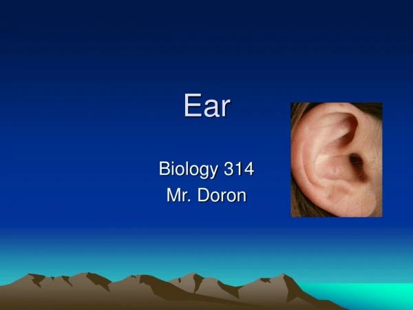
EAR
EAR. EAR OBJECTIVES. Know the microscopic anatomy and histology of inner, outer, and middle ear. Enabling Objectives Describe the components of the outer ear . Discuss the nature of cartilage found in the pinna . Name the parts of the auricle .

EAR
E N D
Presentation Transcript
EAR OBJECTIVES • Know the microscopic anatomy and histology of inner, outer, and middle ear. • Enabling Objectives • Describe the components of the outer ear. • Discuss the nature of cartilage found in the pinna. • Name the parts of the auricle. • Discuss the strata of the tympanic membrane. • List the spaces of the middle ear and what other spaces with which they are continuous. • Describe the ear ossicles and indicate mechanisms whereby sound is mechanically transferred and magnified across middle ear cavity. • Discuss the difference between the membranous and osseous portions of inner ear.
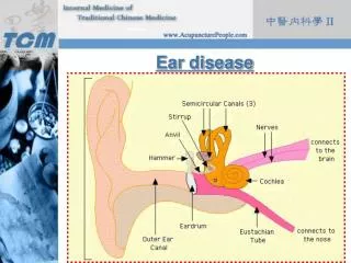
EXTERNAL EAR (Air filled cavity) • Auricle (Pinna) • Typical thin skin with hair and sebaceous glands • Irregularly shaped internal plate of elastic cartilage • Function: • Soundlocalization • Sound amplification
EXTERNAL ACOUSTIC MEATUS • The external acoustic meatus is an air-filledslightly S-shaped tubular space. • It course for about 25 mm to the tympanic membrane (eardrum). • External Acoustic Meatus compose to: • Thin Skin: • Stratified Squamous Epithelium: Epitheliumchanges from keratinized to non- keratinized near the tympanic membrane. • Large sebaceous glands • Ceruminous Gland: Apocrine Coiled Tubular Modified Sweat glands. • Outer one of third is supported by Elastic Cartilage. • The inner two-thirds is formed by Temporal Bone.
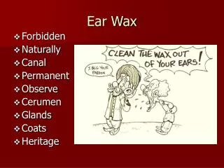
CERUMEN (EAR WAX) • Cerumen (ear wax):Mixture of ceruminous and sebaceous secretions and desquamated meatal cells produces thick waxy product called cerumen or ear wax.
Tympanic membrane separates from external ear to tympanic cavity of middle ear. Tympanic membrane is composed to four strata arranged in 3 general layers: Outermost (cutaneous - ectoderm);very thin skin. Middle (connective tissue - mesoderm): Radiate (radiatum) fibroelastic connective tissue Circular (circulare) fibroelastic connective tissue Innermost(mucosum - endoderm);with low cuboidal epithelium This epithelium continuous with lining of tympanic cavity. TYMPANIC MEMBRANE
TYMPANIC MEMBRANE • Tympanic membrane, close to attachment of manubrium of malleus. • Epidermis (stratified squamous epithelium); • Fibrous layer/lamina propria (consisting of 3, 4, 5); • Network of elastic fibers in subepidermal connective tissue layer; • Outer radial layer of collagen fibers; • Inner circular layer of collagen fibers; • Simple low cuboidalepithelium of tympanic cavity.
TYMPANIC MEMBRANE • The membrane is divided into 2 parts: • pars tensa: • This is the lower four-fifths of the tympanic membrane. • It contains an organized core of connective tissue. • pars flaccida: • This is the upper one-fifth of the membrane. • It lacks a middle fibrous layer and is more flexible.
MIDDLE EAR • The primary function of the middle ear is to convert sound waves (air vibrationsintomechanical vibrations that are transmitted to the inner ear. • The middle ear is an air-filled space in the temporal bone called the tympanic cavity. • It is bridged by three small bones, the auditory ossicles, • They are connected by two moveable joints. • The middle ear includes two openning in the medial wall (vesitubular-oval and cochlear -round window. • The middle ear also contains the internal auditory canal (the Eustachian canal).
Histology of the Tympanic Cavity • Tympanic membrane with malleus, • Epithelium of inner external auditory meatus. • temporal bone; • tympanic membrane (thicker at the attachment site); • malleus (simple squamous epithelium covering bone). • The tympanic cavity is an air-filled space lying within the temporal bone. • It is lined by simple squamous to low cuboidal epithelium. • These cells become ciliated and cuboidal near the opening of the auditory tube. • The lateral wall is composed of the tympanic membrane; • Themedial wall is shared by the inner ear.
OSSICLES OF MIDDLE EAR • The three small bones known as the ossicles, • The malleus (hammer), • The incus (anvil), • The stapes (stirrup), • Cross the space of the middle ear in series and connect the tympanic membrane to the oval window. • One of the ossicles, the malleus, is attached to the tympanic membrane. • The incus links the malleus to the stapes, • The stapes fits into the ovalwindow leading to the inner ear.
HISTOLOGY OF OSSICLES • They are composed of compact bone • These bones are joinedto each other by synovial joints. • The ossicles are covered by epithelium that is continuous with the epithelium of the tympanic cavity. • Function of Ossicles: • Sound in the form of pressure waves causes the tympanic membrane to vibrate, and these vibrations are transmitted to the attached auditory ossicles. • Ossicles provideto link from the external ear to the inner ear.
MUSCLES OF MIDDLE EAR • Muscles attach to the ossicles and affect their movement. • The tendon of the tensor tympani inserts on the malleus. • Contraction of this muscle increases tension on the tympanic membrane. • The stapedius tendon inserts on the stapes. • Contraction of the stapediusreducesthe movement of the stapes at the oval window. • The stapedius, only a few millimeters in length, is the smallest of all the skeletal muscles. Tensor tympani, 40X.(1) Tympanic membrane; (2) malleus; (3) tendon of tensor tympani; (4) tensor tympani muscle; (5) temporal bone.
FUNCTION OF MIDDLE EAR MUSCLE • The two muscles of the middle ear are responsible for a protective reflex called the attenuation reflex. • Contraction of these muscles makes the chain of ossicles more rigid, thus reducing the transmission of vibrations to the inner ear. • This protects the inner ear from the damaging effects of very loud sound. • By contracting, these muscles also improve auditory discrimination by the attenuation of the response of the auditory system to loud background noise
AUDITORY (EUSTACHIAN) TUBE • The auditory (Eustachian) tube is a narrow flattened channel, approximately 3.5 cm long. • This tube is lined with ciliatedpseudostratifiedcolumnar epithelium, about onefifth of which is composed of goblet cells. • It vents the middleear, equalizing the pressure of the middle ear with atmospheric pressure. • The walls of the tube are normallypressed together but separate during yawning and swallowing.
CLINICAL CORRELATION: OTITIS MEDIA • The Eustachian tube runs from the middle of each ear to the back of the throat. • This tube drains fluid normally made in the middle ear. • If the Eustachian tube becomes blocked, fluid can build up. • This can lead to infection. • Ear infections are common in infants and children, because the Eustachian tubes become easily blocked. • Symptoms: Irritability and Crying (infant), Ear pain, fullness in the ear.
THE INNER EAR • Inner ear is located within the petrous portion of the temporal bone. • There are three components to the inner ear: • cochlea, • vestibule, • semicircular canals
THE COCHLEA • The cochlea is a spiral channel that makes about 2-3/4 turns around a central bony core called the modiolus. • The inside of the cochlea (the lumen) is continuous with that of the vestibule. • It connects to the vestibule on the side opposite the semicircular canals.
THE COCHLEA • The cochlea is divided into three spaces by twomembranes: • Membranes: • TheVesitibular (Reissner's) membrane • TheBasilar membrane. • Spaces: • Between these two membranes is the cochlear duct which is also called the scala media. • The cochlear duct is filled with a fluid called endolymph. • The Scalavestibuli • The Scalatympani
THE COCHLEA • At the basal end of the scalavestibuli is the oval window, which is connected to the auditory ossicles that transmit vibration from the eardrum. • At the basal end of the scala tympani is the membrane-covered round window, which absorbs or dampens vibrations reaching it, thereby providing a compensatory release of the vibratory pressures at the oval window.
ORGAN OF CORTI • The Organ of Corti is the sensor of sound vibrations. • The Corti organ is locatedwithin the cochlear duct (scala media). • The upper wall of the cochlear duct is the Vesitubular (Reissner's)membrane • Thelower wall or floor of the cochlear duct is the Basilarmembrane. • The organ of Cortirests on the basilar membrane and is overlain by the tectorialmembrane.
HISTOLOGY OF ORGAN OF CORTI • The organ of Corti is composed of hair cells and supporting cells. • The inner hair cells, about 3500 in number, are arranged in a single row on the inner side of the inner rods of Corti. • The 12000 outer hair cells are longer, and are arranged in three rows in the basalcoil of the cochlea, • Outer hair cells are organized in four or fiverows in the apical coil of cohlea.
HEARING • Sound waves hit on the tympanic membrane • Tympanic membrane translates into simple mechanical vibrations. • The ossicles of the middle ear convey these vibrations to the cochlea. • Movement of the stapesis found on the oval window. • The vestibule collections vibrations from oval window. • Travelling wavestransfer in the perilymph of the vestibular canal. • The vibrations are transmitted through the vestibular membrane to the cochlear duct, which contains endolymph.
HEARING • Vibrations that spread through the cochlea induce vibrations in the basilar membrane, • Then vibration is transduced into afferent nerve excitation by the hair cells. • Each hair cell is covered with 50 to 100 hairlikestereocilia which are modified microvilli. • Mechanical vibration hits the stereocilia of hair cell and then stereocilia is bended. • Stretching of the plasma membrane caused by bending of the stereocilia. • Hair cell generates membrane potential changes in the receptor cell.
HEARING • Signals are conveyed to the afferent nerve endings associated with each hair cell. • Bending of stereociliamake a depolarization of hair cells • Then stereocilia bends the bending in the opposite direction and produces hyperpolarization.
THE VESITUBULER APPARATUS • CochlearDuct consists of fivevestibular structures. • Eachstructures contains a specialized mechanoreceptor: • Maculae within the utricle and saccule, • Cristae within the ampullae of the superior, horizontal, and posterior semicircular canals.
The saclike utricle and saccule contain areas called maculae,. They specifically respond to linear acceleration, such as the pull of gravity. The threesemicircularcanals within swellings, this is called ampullaeand swellings containtheCristae Crista is the specialized detectors of angular acceleration. THE VESITUBULER APPARATUS
The vestibular labyrinth receives a dual innervation: Thedistal axonal processes of vestibular afferent neurons, whose cell bodies are in the vestibular ganglion, The vestibular efferent fibers that originate in the brainstem. The afferent axons terminate on the mechanoreceptivevestibularhair cells that are the basic sensory transducers of the labyrinth. THE VESITUBULER APPARATUS
CRISTAE • Cristae include two type hair cell; type I and type II. • Type I hair cells are thought to be more sensitive than those of type II. • The apical ends of both types of hair cells bear a tuft of 40 or more sensory hairs, or stereocilia, • The sensory hairs of the mechanoreceptors in the cristae are embedded in the gelatinous cupula • Cupula extends to the opposite wall of the ampulla.
The maculae of the utricle and saccule are oriented at right angles to one another. When a person is standing; The macula utriculi is in a horizontal plane, The macula sacculi is in a vertical plane. Each macule consists of hair cells and nerve endings associated with the hair cells. The gelatinous material that overlies each macula is called the otolithic membrane. It contains tiny crystalline particles of calcium carbonate and protein, called otoliths (otoconia). Theotoconia are denser than the surrounding fluid, the otolithic membrane tends to move under the influence of linear acceleration. MACULAE
The Role of Maculae in Maintaining Equilibrium • Linear acceleration: Movement of the head in a linear direction causes the otolithicmembrane to slide backward or forward over the hair cells, bending the hairs. • When the hair cells are bent toward the kinocilium, the hair cells depolarize and impulses sent to the brain increase in frequence. • When the hairs are bent in the opposite direction, the receptors hyperpolarize and impulse generation declines. • Note that the maculae respond only to changes in acceleration or velocity of head movement.
The Role of Cristae in Maintaining Equilibrium • Rotational (angular) acceleration: Thethree semicircular canals - superior, posterior, and horizontal - are positioned at right angles to one another, • They are able to detect movements in three-dimensional space. • During rotational movement of thehead, the walls of the semicircular canal and the membranoussemicircular ducts move, • Butthe endolymphcontainedwithin the ducts tends to lag behind because of inertia. • Thecupula, projecting into the endolymph, is swayed by themovement differential between the crista fixed to the wall ofthe duct and the endolymph. • The deflection of the cristae excites the hair cells by bending the cilia. • Deflection in one direction depolarizes the cells; • Deflectionin the other direction hyperpolarizes them.
CRISTAE AMPULLARIS • Cupula; • Sensory epithelium (with stereocilia); • Transitional epithelium (no "hairs"); • Dark cells (melanin underneath); • Lamina propria with blood vessels, myelinated nerve fibers
MACULAE • Sensory epithelium; • Remnants of otholithic membrane; • order cells; • Vestibular nerve fibers; • Temporal bone.