EAR
EAR. EAR. EAR IS THE SENSE ORGAN WHICH HELPS US IN HEARING IT ALSO HELPS US IN MAINTAIN THE BALANCE OR EQUILIBRIUMOF OUR BODY COCHLEA IS THE MAIN PARTS WHICH HELPS IN HEARING. PARTS OF THE EAR. EXTERNAL EAR. INNER EAR. MIDDLE EAR. EXTERNAL EAR.

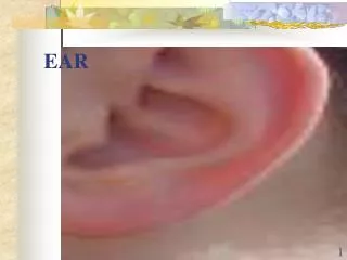
EAR
E N D
Presentation Transcript
EAR • EAR IS THE SENSE ORGAN WHICH HELPS US IN HEARING IT ALSO HELPS US IN MAINTAIN THE BALANCE OR EQUILIBRIUMOF OUR BODY • COCHLEA IS THE MAIN PARTS WHICH HELPS IN HEARING
PARTS OF THE EAR EXTERNAL EAR INNER EAR MIDDLE EAR
EXTERNAL EAR • EXTERNAL EAR CONSISTS OF PINNA AUDITORY CANAL AND EAR DRUM .PINNA HELPS TO DIRECT THE SOUND WAVESINTO THE AUDITOR CANAL. • CERUMINOUS GLAND IS SPECIAL GLAND WHICH ARE FOUND IN THE WALLS OF THE AUDITOURY CANALWHICH IS THE CONSTITUTION OF PINNA. THE WAX PRODUSED BY THESE GLANDS AND THE HAIR IN THE AUDITORY CANAL TOGETHER PROTECT THE EAR FROM SMALL INSECTS, GERMS AND DUST.IT HELPS IN MAINTAIN THE TEMPERATURE AND DAMPNESS OF THE AUDITORY CANAL ENDS IN THE EAR DRUM.
MIDDLE EAR • MIDDLE EAR IS AN AIR FILLED CAVITY WHICH LIES • JUST BEYOND THE EAR DRUM. • MIDDLE EAR CONSIST OF 3 SMALL BONES • 1)HAMER SHAPED BONE ATTACHED TO EAR DRUM • IS CALLED MALLEUS • 2)ANVIL SHAPED BONE IS CALLED INCUS • 3)THIRD BONE IS THE STAPES IT IS THE SMALLEST • BONE IN THE HUMAN BODY. • AN AIR PASSEGE CALLED EUSTACHIAN TUBE HELPS TO CONNECT THE MIDDLE EAR WITH PHARENIX.EUSTACHIAN TUBE HELPS TO MAINTAIN AIR PREEASURE BALANCE OF EITHER SIDE OF EAR DRUM
INNER EAR • INNER EAR IS SITUATED IN THE BONY CAVITY IT CONTIN 2 LABRINTH • 1)BONY LABRINTH • 2)MEMBERANEOUS LABRINTH • INNER EAR IS MADE OF 2 PARTS WESTIBULAR APPARETUS AND CHOCLEA IT INCLUDES 3 SEMICIRCULARCANALS. • EACH SEMICIRCULAR CANAL THERE IS A SWELLING CALLED AMPULLA CONTAIN RESIPTORS CALLED CRISTATE UTRICLE AND SACULES CONTAIN RESIPTORS AND MACULAE
CAUSES OF DEAFNESS • INFECTION IN THE EUSTACHIAN TUBE WILL SPREAD TO MIDDLE EAR TOO • EAR DRUM MAY BECOME DAMAGED BY AN INFECTION IN THE AUDITORY CANAL • LEADING TO DEAFNESS IS EXCESS NIICE STRONG BLOW ON THE CHEAK • POINTED OBJECT ENTERING THE EAR • ATTACK OF THE INSECT LEAD DEAFNESSDEFECT IN THE ,CHOCLEA,AUDITORY CANAL,BRIANARE ALSO REASON FOR DEAFNES
This is an animation of a bird's hearing system. It is very similar to the human equivalent, except that the cochlea (on the right) doesn't spiral in so many times. The bird cochlea has 0.75 turns; The human cochlea has 2.5 turns
On the far left is the eardrum. This vibrates in response to incoming sound. From the eardrum, the vibration passes along the three bones, the hammer (which is connected to the eardrum), the anvil and the stirrup. It is not too easy to see in this diagram, but the bones are similar in shape to the items after which they are named. • The last of the bones, the stirrup, moves with about 20 times the force of the hammer due to the lever principal. So these bones serve as a form of amplification. • From the stirrup, the vibration passes through the oval window into the cochlea, where it causes a liquid called the cochlear fluid, located in the upper and lower galleries of the cochlea (see later diagram) to vibrate. In between the two galleries, is the organ of corti, which spirals along the length of the cochlea (shown as a black and yellow line in the first drawing).
It is inside the organ of corti that we find the hair cells.These are nothing to do with hairs on your head. They are the receptors involved in changing the sound pressure wave into a nerve signal. Hair cells get their name from the hair like projections at their top which are known as 'cilia'. There are 50 to 100 'hairs' on each hair cell.
Here is a cutaway diagram of the human cochlea seen from a different perspective. It is in all about 1.5 cm in diameter. You will see that the spiralling tunnel has a bony shelf that divides it into two galleries; the upper and the lower. The organ of corti runs along the edge of this bony shelf. • different positions along the length of the organ of corti inside thecochlea respond to different frequencies.
The organ of corti, which runs the whole length of the cochlea, contains two subtly different types of hair cells; the inner hair cells (IHCs) (green in the diagram)and the outer hair cells (OHCs) (blue).Current theory (Which is not 100% perfect, but is 'state of the art') tells us that the IHCs are in the main responsible for the actual 'hearing' of sounds, while the OHCs, which are really tiny muscles, are responsible for 'fine tuning' the frequency response - sharpening up that narrow notch mentioned earlier. • This diagram shows only a small section of the structure. It actually runs the whole length of the coiled cochlea. The hairs, or cilia ,on the inner hair cells which detect the sound are shown in green.The hairs on the outer hair cells are shown in blue. The cells themselves are underneath. The dark circle in the middle of each cell is its nucleus. Note that there are three rows of outer hair cells and just one row of inner hair cells
TO PROTECT EAR • AVOID SWIMMING IN CONTAMINATED WATER. • DONOT INSERT SHARP OBIECT INTO THE EAR IT CAN CAUSE INJURY • AVOID HIGH FREQUENCY SOUND • NEVER STRIKE ANY ONE ON THE CHEAK EVEN FOR FUN • AVOID INFECTION IN THE INTERNAL EAR
Prepared by • Jithin krishna.R (X A) • Arjun Manoj (X A) • Sooraj Dev.J.R (X A) • Mohammed Suhail (X D) • S.M.V.H.S.S