Download
1 / 69
690 likes | 863 Views
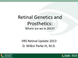
Retinal Genetics and Prosthetics: Where are we in 2013?. VRS Retinal Update 2013 D. Wilkin Parke III, M.D. Objectives. Describe the clinical value of current genetic testing for AMD Describe some currently available retinal prostheses and clinical scenarios in which they might be beneficial.

E N D
Retinal Genetics and Prosthetics:Where are we in 2013? VRS Retinal Update 2013 D. Wilkin Parke III, M.D.
Objectives • Describe the clinical value of current genetic testing for AMD • Describe some currently available retinal prostheses and clinical scenarios in which they might be beneficial
Scenario 1 • You’re on a flight out of town and the guy next to you recognizes you as his mother’s doctor • Mom has AMD and son desperately wants to know whether the whole family should get genetic testing • You blame the ad for an AMD gene test that you see in the in-flight magazine • Your smart phone is turned off and it’s a three-hour flight • What do you say?
Genetic testing for AMD #1: What role do genes play in development of AMD and advanced AMD? #2: Which genes look like the big players? #3: Can we risk stratify patients yet? • Is this any better than a good exam? #4: Can we target therapy to genotype?
AMD in the U.S. • 2020 • 3 million with AMD • 400,000 with wet AMD 2012 • 2.2 million with AMD • 300,000 with wet AMD • Not only is it a leading cause of blindness, but 50% of all new registered blindness! • 30% greater than 75 will have it
Risk Factors Modifiable: • Smoking • Hypertension • Hyperlipidemia • Obesity • Sunlight exposure Not modifiable: • Genetics • Age
Genetic testing for AMD #1: What role do genes play in development of AMD and advanced AMD? #2: Which genes look like the big players? #3: Can we risk stratify patients yet? • Is this any better than a good exam? #4: Can we target therapy to genotype?
How important are genes in AMD? • FH: First degree relative is at 6-12x higher risk than the general population • Genetic variants are responsible for 60-70% of the risk (Seddon et al 2009, Spencer et al 2011)
AMD Gene Consortium • Confirmed 12 and identified 6 more loci of AMD “susceptibility” in a meta-analysis of 7600 cases (Holliday et al 2013) • Asian and European gene markers appear different in prevalence and significance
Genetic testing for AMD #1: What role do genes play in development of AMD and advanced AMD? #2: Which genes look like the big players? #3: Can we risk stratify patients yet? • Is this any better than a good exam? #4: Can we target therapy to genotype?
Genes in AMD • Complement factor H (CFH) • Chrom 1q31 • The first one for AMD, 2005 • Alternate complement pathway • ARMS2/HTRA1 • Chrom 10q26 • Age-related maculopathy susceptibility factor 2 • Extracellular matrix and basement membrane formation • Others: • Chromosome 6 • Complement component 2 (C2) and complement factor B (CFB) • Nearby genes for VEGF-A and Col10A • Chromosome 9 • Nearby genes for Col15A1, TGFBR1, ABCA1 • Weaker associations on chromosomes 2, 3, 4, 5, 8, 12, 15, 17, 18, 21
Rare variants • CFH mutation (CFHR1*B) associated with hemolytic uremic syndrome, • found in some individuals with nonsyndromic AMD • PRPH2 gene mutation is associated with a CACD-like macular atrophy • ABCA gene polymorphisms have been associated with severe AMD
Rare variants (cont’d) • Elastin mutations identified in Japanese with AMD • Leads some to think that IPCV may be a subtype of AMD expressed in certain genetic variations • Case control studies are not possible with these conditions—they’re too rare • Distinguishing atypical AMD from other macular diseases can be difficult
Genes to remember • ARMS2 • Chromosome 10 • CFH • Chromosome 1
What role do genes play in development of AMD and advanced AMD? • Probably a large one, but there are too many contributing genes and too much environmental modification for us to categorize it as predominantly inherited • Which genes look like the big players? • CFH and ARMS2 on chromosomes 1 and 10.
Genetic testing for AMD #1: What role do genes play in development of AMD and advanced AMD? #2: Which genes look like the big players? #3: Can we risk stratify patients yet? • Is this any better than a good exam? #4: Can we target therapy to genotype?
Talking about odds ratios • Characteristic 1q31 and 10q26 variants have the strongest association with development of advanced AMD • But even for these, odds ratios are difficult to define • Ratios vary based on the study • Different populations • Different phenotypic characteristics • Almost all are case control studies—not true measurements of relative risk
Odds ratios for high risk genotypes • CFH (Y402H variant) • Odds ratio of 2-2.5 in Europeans • ARMS2 • Odds ratio of 6-10 for highest risk genotype • C2/CFB • Protective alleles may reduce risk by 45-53% • CFH and ARMS2 – highest risk genotypes for both • Odds ratio of 62 • Smoking – 10-15% current population, • Odds ratio of 2.5-6 • But these are compared to “normal” age-matched controls! • This is not from prospective monitoring of a population as it ages
What we know • Those with the highest concentration of high risk alleles have a higher risk than those with the lowest concentration of high risk alleles • Most patients are in the middle ground • Most authors agree current tests lack “the level of sensitivity and specificity that one would normally demand of a clinical test” (Jakobsdottir et al 2009)
Clinical severity score In each eye: • 1 point for presence of large drusen • 1 point for presence of pigment epithelial abnormality Ferris FL et al. A simplified severity scale for age-related macular degeneration: AREDS Report No. 18. Arch Ophthalmol2005; 123(11):1570-4.
Clinical severity score Further modification by age, smoking status, family history www.ohsucasey.com/amdcalculator Ferris FL et al. A simplified severity scale for age-related macular degeneration: AREDS Report No. 18. Arch Ophthalmol 2005; 123(11):1570-4.
Clinical severity score Factoring in the CFH and ARMS2 variants changes the score, but not by much Ferris FL et al. A simplified severity scale for age-related macular degeneration: AREDS Report No. 18. Arch Ophthalmol2005; 123(11):1570-4.
So is this any better than a good exam? • Probably not, at least right now
Genetic testing for AMD #1: What role do genes play in development of AMD and advanced AMD? #2: Which genes look like the big players? #3: Can we risk stratify patients yet? • Is this any better than a good exam? #4: Can we target therapy to genotype?
The only things to reduce risk • Stop smoking • Low glycemic index diet • Lutein and zeaxanthin • Vitamin D (only to avoid deficiency) • Beta-carotene, zinc • UV protection
Vitamins and genotype • Antioxidants, lutein, zeaxanthin, and zinc might reduce impact of high risk genotypes (Ho et al 2011, Klein et al 2008)
Anti-VEGF therapy and genotype • One homozygous CFH genotype and one VEGFA gene variant may be predictive of improved response to anti-VEGF (Chen et al 2012, Abedi F et al 2013) • No consistent evidence yet of association between at-risk alleles on chromosomes 1 and 10 and either positive or negative responders to therapy (Orlin et al 2012)
Alternative Screening • Home-based monitoring in the near future • iPhone app • Foresee Home device • Can we tailor the intensity of home screening to genetic risk? www.foreseehome.com www.digisight.com
Patient motivation • Testing early might motivate higher risk individuals to address risk factors more aggressively • But this could disadvantage lower risk patients. It might produce surprise and disillusionment if they still get advanced AMD • Genetic testing is rarely straightforward
Genetic testing:The holy grail • In the future we may find the risk-benefit balance for each age, clarify the pharmacogenetic associations, and develop specific monitoring and therapy.
“Avoid routine genetic testing for genetically complex disorders like age-related macular degeneration and late-onset primary open angle glaucoma until specific treatment or surveillance strategies have been shown in 1 or more published clinical trials to be of benefit to individuals with specific disease-associated genotypes” (Stone et al 2012)
Scenario 2 • You had a great vacation and you’re on the flight home • The flight attendant overhears what you do • Her son has RP and she’s saving up to send him to Italy for a retinal prosthesis • She’s happy to take your drink order if you’ll only tell her whether the prosthesis is worth it • Your smart phone is turned off and it’s still a three-hour flight • What do you say?
Retinal prosthetics #1: How do they work? #2: What types are available, and when are these being used right now? #3: With which patients do we have this discussion?
Retinal prosthetics #1: How do they work? #2: What types are available, and when are these being used right now? #3: With which patients do we have this discussion?
Retinal prosthetics:The rationale • Outer retinal disorders • Postmortem analyses indicate that after total photoreceptor loss in RP, that up to 90% of inner retinal neurons can remain histologically intact. • The visual pathway downstream to the photoreceptors remains theoretically viable
Retinal prosthetics • Electronic implants • Non-electronic implants
Retinal prosthetics • Electronic implants • Non-electronic implants
The parts • Encoder – converts light into electrical energy (retina’s data) • Transducer implant • Formulates stimulation pattern • Triggers electrodes • Electrodes fire in close proximity to target cells (usually ganglion cells) • Target cells activated by proximal electrical charge Weilandet al, 2011
Retinal prosthetics #1: How do they work? #2: What types are available, and when are these being used right now? #3: With which patients do we have this discussion?
Epiretinal Prosthesis Epiretinal implant/electrode array Extraocular receiver Wireless transmitter Camera in glass frame Humayun, et al (2003)
Epiretinal Prosthesis 4x4 platinum electrode array Humayun, et al (2003)
Epiretinal Prosthesis • Yanai (2007): • Visual performance tested via simple visual tasks: • Locate and count objects • Differentiate three objects • Determine orientation of a capital L • Differentiate four directions of a moving object • Performance was significantly better than chance in 83% of the tests
Subretinal Prosthesis Chow, et al (2004)
Subretinal Prosthesis • Chow AY, Pollack JS et al (2004): • Silicon-based subretinal microchip • 5000 microelectrode-tipped microphotodiodes powered by incident light • Implanted subretinally in 6 patients • Subjective visual improvement seen in all patients
Problems and limitations with electronic prostheses • Power: • Large heat dissipation per electrode • Implants can’t heat tissue more than 1 degree Celsius. • Limits electrode number • Cochlear implants do well with only 16 electrodes, but vision requires more resolution • Triggering the appropriate “on” and “off” neurons • The inner retinal layers show some architectural and functional change with the photoreceptor degeneration, so the downstream system may not be “normal”
Retinal Prostheses and RP Modified from Weiland, et al, 2011
Collective experience • Since 2002, published series with retinal prostheses have come out of the US, Italy, France, Germany, the UK, and Japan. • The first clinically approved Argus II was performed 10/2011 in Italy. • The Argus II received FDA approval for adult advanced RP on February 13, 2013. • Over 70 patients with end-stage RP have received one.
Retinal prosthetics #1: How do they work? #2: What types are available, and when are these being used right now? #3: With which patients do we have this discussion?