Download
1 / 44
450 likes | 763 Views
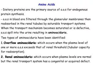
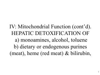
IV: Mitochondrial Function (cont’d). HEPATIC DETOXIFICATION OF a) monoamines, alcohol, toluene b) dietary or endogenous purines (meat), heme (red meat) & bilirubin,. Detoxification of monoamines catalysed by mitochondrial outer membrane MAO. e.g., dopamine norepinephrine tyramine

E N D
IV: Mitochondrial Function (cont’d). HEPATIC DETOXIFICATION OF a) monoamines, alcohol, toluene b) dietary or endogenous purines (meat), heme (red meat) & bilirubin,
Detoxification of monoamines catalysed by mitochondrial outer membrane MAO e.g., dopamine norepinephrine tyramine phenethylamine octylamine serotonin outer membrane MONOAMINE OXIDASE Flavin-containing amine oxidase R-CHNH2 inner membrane R-CH=NH (imine) O2 NH3 H2O2 R-CHO NAD+ ATP, HCO3- aldehyde dehydrogenase urea cycle UREA NADH R-COOH b-oxidation CO2 Mitochondrion
Amine specificity for the two isoforms (A & B) in humans MAO A preferentially metabolizes serotonin. MAO B preferentially metabolizes phenethylamine, dopamine. MAO inhibitor + dietary amines or proprietary drugs MAOB inhibitor deprenyl (selegiline) similar to phenethylamine and increases brain dopamine levels. This is used to treat Parkinson’s disease. But “Hypertensive crisis” is a hyperadrenergic state induced by MAO inhibitors + pressor amines (e.g., tyramine in cheese,beer,wine or soya sauce) or proprietary drugs (e.g., L-DOPA, mazindol, ephedrine, etc). Perry, 1996. http://www.vh.org/adult/provider/psychiatry/CPS/19.html
Detoxification of alcohols by matrix ALDH2 Covalently binds to protein-NH2 (ADH) cytosolic alcohol dehydrogenase acetaldehyde ETHANOL ANTABUSE (disulfiram) or cyanamide NADH NAD+ Methyl pyrazole NAD+ aldehyde dehydrogenase CO2 NADH (ALDH) TCA cycle ATP acetate acetylCoA CoASH
COOH URINE CONHCH2COOH Hippuric acid c) Benzoic acidosis induced by toluene glue sniffing Benzaldehyde Toluene Benzylalcohol ADH Cyt P-450 CH3 CH2OH CHO CYTOSOL ER ALDH1/ ALDH2 MITOCHONDRIA Benzoic acid Benzoyl-CoA synthetase ATP CoA N-acyltransferase Benzoyl-CoA glycine 5 Teratogen Update: Toluene teratology. 55, 145-51, (1997)
8. Hemoprotein toxicity diseases caused by heme or bilirubin A) Rhabdomyolysis (drug induced) releases myoglobin and heme and causes kidney mitochondrial damage B) Kernicterus:- bilirubin causes neonatal brain mitochondrial damage
A) Myoglobin mediated rhabdomyolysis rarely caused by statins STATIN Drug BAYCOL withdrawn by BAYER, 2001 CRUSH INJURY Or if LAIN IMMOBILE for hrs (e.g. EtOH, heat stroke) Muscle Cell Drug reactive metabolites Mitochondrial/cytotoxicity • x10 creatine • phosphokinase • Dark brown granular • casts in urine Myoglobin release Massive muscle breakdown Acid Mb Fe accumulates in kidney renal tubule Acid lipid peroxidation (proximal tubule) mitochondrial toxicity KIDNEY Failure (7% of all cases of acute renal failure) Spontaneous myalgias Muscle tenderness, Weakness, malaise, fever DEATH THERAPY 1.) NaHCO3 base 2.) Desferoxamine CMAJ 165(5) pg 632 (2001); J. Biol. Chem. 273, 31731-7 (1998)
B) Hemoglobin/heme mediated brain damage ie. Kernicterus (brain damage caused by bilirubin in the newborn (hyperbilirubinemia)) • re-emergence (due to shorter hospital time, breastfeeding) • danger signs onset of jaundice 3 days following birth, vomiting, lethargy, poor • feeding, fever, high pitched crying. • plasma bilirubin > 340 uM = exchange transfusion therapy • plasma bilirubin > 240 uM = Phototherapy (blue light) • Drug therapy Sn protoporphyrin (inhibits heme oxygenase). • Hemoglobin bilirubin • Bilirubin binds to released brain mitochondrial • serum albumin by competing toxicity (hearing • fatty acids or loss? brain damage • drugs encephalopathy) Heme oxygenase of reticuloendothelial cells of spleen, liver, kidney Glucuronyl Transferase Appears at 7d after birth Glucuronide conjugated bilirubin. • 0.3% mortality (yellow staining of brain) • family history, hemolytic diseases, Gilbert’s disease, diabetes (erythrocyte fragility), • G6PD deficiency (Pediatrics 106, 1478-80 (2000), sickle cell.
Metabolic pathway of heme degradation to bilirubin and detoxification of bilirubin by glucuronidation. (M=methyl,V=vinyl and P=propionate represent heme side chains CO Fe2+ Heme Oxygenase NADPH: P450 Reductase NADPH NADP+ NADPH O2 NADP+ H2O NADPH NADPH Biliverdin Reductase NADP+ UDP UDP-glucuronic acid UDP-glucuronosyl transferase JAUNDICE Antioxidant, but at high concn. TOXIC TO NEONATAL BRAIN MITOCHONDRIA
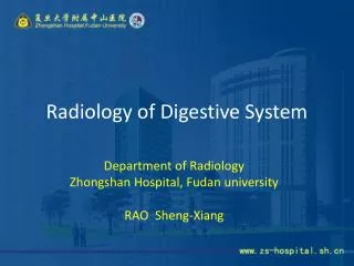
9. HEME BIOSYNTHESIS BYMITOCHONDRIA AND GENETIC DISEASES A) Heme biosynthesis for cytochromes, etc.B) Genetic diseases: Porphyrias and porphyrin toxicityC) Oxidative degradation of heme to bilirubin
Heme synthesis • Heme required for synthesis of mitochondrial cytochromes and endoplasmic reticular P450s. • Heme required for bone marrow synthesis of hemoglobin and muscle myoglobin • STEP 1 for heme synthesis is the synthesis of aminolevulinic acid (ALA) from succinyl CoA of the citric acid cycle and glycine
Heme biosynthesis in bone marrow (hemoglobin) and liver (cytochromes) NH2 or lead 1
Erythropoietin synthesised by kidney induces hemoglobin synthesis in bone marrow (replaces blood transfusion!)
Heme Biosynthesis - STEP 3 - mitochondrial final steps (3 enzymes)
B) PORPHYRIAS I. Enzymes Inhibitors causing porphyria II. Genetic porphyrias
I. ENZYME INHIBITORS CAUSING PORPHYRIA PORPHYRIA - Greek “porphyros” (purple urine) an over-production disease. Often neuropsychiatric (syphilis much more so). 2) ALA dehydratase - lead 5) Uro’gen decarboxylase - PCBs, dioxin, lead, cadmium, hexachlorobenzene (fungicide - seeds). Mitochondrial oxidase - steroids (birth control pills) and estrogens 8) Ferrochelatase – lead II. GENETIC PORPHYRIAS (Emerg Med Clin.N Am 23 (2005)885-899) 2) ALAD deficiency porphyria (Seminars in Liver Disease, 18, 95-101, 1998) excrete ALA not PBG, symptoms like (neurological).
Scriber et al. (eds). Metabolic and molecular basis of inherited disease. 7th ed. 1995, p.2124.
3) Acute intermittent porphyria (i.e. phosphobilinogenin deaminase deficiency) excrete ALA and PBG , the most common hepatic porphyria)- MEDALERT bracelet • 1. Symptoms - excrete PBG and ALA in the urine , high plasma PBG/ALA • - severe abdominal pain, nausea, vomiting, hypertension(cardiovascular) • then CNS - anxiety, insomnia, confusion, hallucination, paranoia then • peripheral neuropathy - fatal respiratory paralysis. Liver cancer. • 2. a. PBG deaminase deficiency - gene locus 11q24 (>100 mutations), autosomal • dominant 1:10,000-50,000 but 1:1000 in N. Sweden (Lapland) • b. Precipitated by phenytoin, phenobarbital drugs, alcohol, fasting, • hormones estrogen, stress, infection or lead poisoning • 3. Biochemistry of neurotoxicity • a. heme deficiency P450 drug metabolism tryptophan dioxygenase • brain tryptophan brain serotonin mitochondrial cytochromes (mitoch.disease) • b. ALA “ROS”, mitochondrial DNA damage. • 4. Therapy –ALA with heme arginate (taken up by liver not b.m.), high glucose • 5. Mouse model (PBG knockout) - called Vincent! - behavioural studies • Suspects - Van Gogh, King George III(?) (Absinthe for insomnia) • 6.Diagnosis :- Patients urine exposure to sunlight (reddish brown fluorescence)
4) Congenital erythropoietic porphyria (Erythroid, Photosensitive) • -Fe overload of b.m., uroporphyrin overload of skin and erythrocytes • hemolysis • 1. Symptoms: skin lesions (light) , risk of infection in early infancy • Anemia (hemolysis) • Teeth reddish brown (fluorescence) • 2. Uroporphyrinogen III synthase deficiency • Uroporphyrinogen I and coproporphyrin I accumulation in the skin, bone marrow, erythrocytes, urine, plasma • 3. Treatment: activated charcoal (oral); b.m. transplant; gene therapy for bone • marrow; blood transfusion to stop erythropoiesis in b.m. • 4. Only 200 diagnosed so far; werewolf, vampires (early childhood sensitive • to light skin blisters, infection disfigures) is not porphyria
5) Porphyria Cutanea Tarda - “heme deficiency and uroporphyrin overload of liver” (Photosensitive; commonest porphyria;readily treated). 1. Symptoms: lesions on backs of hands and face due to photosensitization by uroporphyrin I, III or uroporphyrinogen. Abdominal pain, neural,psychiatric. ALA,porphobilinogen. 2a. Diagnosis :- urinary uroporphyrin >> coproporphyrin. b. Uroporphyrinogen decarboxylase deficiency (liver but also erythrocyte in type II): or hexachlorobenzene or dioxin induced CYP 1A2. (Turkey bread disaster). Also Fe uroporphyrinogen III synthase - uroporphyrin I. c. Candidate hemochromatosis gene leads to diagnosis of hemochromatosis 3. Precip. by alcohol, hepatitis C virus, estrogen (birth control, post-meno- pause, prostate, pregnancy), Fe. 4. Biochemistry - inactivation of decarboxylase (-SH enzyme) by “ROS” from reduced P450/P450 reductase/Fe 5. Therapy:- Fe removal (phlebotomy or desferoxamine and ascorbic acid) - chloroquine endocytosis and releases uroporphyrin chloroquine complex from hepatocytes - heme?
6) Hereditary Coproporphyria - coproporphyrinogen oxidase deficient, chronic fatigue syndrome. - hepatic porphyria Symptoms: liver cancer 7) Porphyria variegata - protoporphyrinogen oxidase 50% deficient. –hepatic and skin porphyria Gene locus 1q2320. 20,000 S. Africans descended from a Dutch lady in 1688 who went to S. African for marriage to another Dutch settler . Now 3/1000 S.Africans have this! Symptoms: skin and acute porphyria if exposed to sunlight,neurovisceral symptoms (abdominal pain),liver cancer Diagnosis :- plasma fluoresces when exposed to UV. Precipitated by sunlight Treatment: increase ALA synthetase by heme arginate, glucose ( NADPH) ?
8) Erythropoietic protoporphyria - associated with liver cancer, red urine “porphyrin and Fe overload disease” “Skin photosensitive” a. Ferrochelatase decreased 75-90% b. Symptoms: photosensitive skin (infancy),velvet knuckles, black liver c. Biochemistry - protoporphyrin IX accumulates in erythrocyte membranes and liver; intramitochondrial Fe accumulates in bone marrow - skin photo- sensitivity results from protoporphyria IX effluxing erythrocytes into plasma. d. Treatment – topical sunscreens and oral beta-carotene (ROS scav.?) e. Diagnosis –incr. protoporphyrin in erythrocytes,plasma,bile,feces I. Liver transplant; operation is difficult as operating room must be kept dark (only yellow acetate filters) II. Abdominal burns; transfused blood is destroyed as blood difficult to store.
C) OXIDATIVE DEGRADATION OF HEME TO BILIRUBIN (a brain neurotoxin in babies)
Detoxification function of the liver a) heme toxin oxidation to bilirubin b) detoxification of bilirubin by glucuronidation or albumin albumin LIVER CO phagosome Heme oxygenase
Degradation of heme STAGE 1 Hemoglobin of old erythrocyte trapped in spleen
STAGE II - ligandin in hepatocyte surface membrane traps bilirubin from plasma and helps transport it into the liver where it is glucuronidated Albumin - bilirubin (a GSH transferase)
*mutated gene in Gaucher’s disease (carried by 15% of population)
Mitochondrial Function (Con’t). a) N-Catabolism of Amino Acids (The Urea Cycle) and the Detoxification of Ammonia (very toxic) • N-Catabolism of Purines • reperfusion injury and drug therapy • Genetic diseases: HGPRT • Gout, Lesch-Nyhan syndrome
Disposal of nitrogen by mitochondria a) The urea cycle: Detoxification of NH3
Mitochondrial Function (Con’t). Formation of urea from amino acids transaminase dehydrogenase Urea cycle dehydrogenase a-ketoglutarate Glutamate
IV: Peroxisomes Function • β-oxidation of fatty acids but oxidase forms H2O2 • Glyceraldehyde is metabolised to D-glycerate and glucose via glycolate (glycolate oxidase forms H2O2 and glyoxylate) • Catalase detoxifies H2O2 to form oxygen and H2O and cooxidises other substrates, including phenols, formic acid, formaldehyde, and methanol. • Glyoxylate detoxified by peroxisomal alanine:glyoxylate transaminase to form glycine & pyruvate. Otherwise glyoxylate would be oxidised by lactate dehydrogenase to oxalic acid which causes oxalate stone formation & kidney damage. • Biosynthesis of bile acids and ether phospholipids
V: CYTOSOLIC FRACTION N-catabolism of purines Glycogen synthesis and glycogenolysis
Disposal of nitrogen (Con’t): b) N-catabolism of purines in the cytosol & peroxisomes HGPRT + PRPP HGPRT + PRPP Voet pg. 714.
Cytosolic Fraction 1) N-catabolism of purines can be lethal Reperfusion Injury e.g. Myocardial infarction or Paraplegia • ISCHEMIC STAGE • Lack of O2no ATP synthesis by heart muscle mitochondria. ADPAMPhypoxanthine • accumulates and leaves muscle cell to enter endothelial cell. • Acidosis causes endothelial cell xanthine dehydrogenasexanthine oxidase • Acidosis results from marked increase in glycolysis to form ATP + lactic acid (to keep cells alive • by maintaining ATP levels) b) Reperfusion of O2 • O2 is the substrate for xanthine oxidase • Damaged endothelial and muscle cells (ischemic toxicity) • release factors which recruit neutrophils (inflammatory reaction activated NADPH oxidaseO2*-. Xanthine oxidase Accumulated xanthine and hypoxanthine O2 (Endothelial cell)
Drugs which prevent reperfusion injury • Allopurinol – inhibitor of xanthine oxidase • Desferal – Complexes Fe2+ • Antioxidants e.g. Lazeroids (upjohn) to prevent membrane lipid • peroxidation • 4) Removal of H2O2 by NADPH (e.g. glucose) or GSH (e.g. methionine) • or dietary Se. H2O2 OH Catalase (activated by NADPH) H2O GSH Peroxidase GSSG + H2O 2 GSH • Anti-inflammatory drugs (e.g. indomethacin or phenylbutazone) • Ca2+ Blockers
Treatment or to prevent Ischemic damage (reperfusion injury)
2) Purine catabolism can be painful and cause joint damage Figure 22.22 (Voet) – By James Gilroy • Defects in excretion of uric acid and/or partial deficiency in HGPRT activity due to mutation in HGPRT gene • Uric acid precipitation in joints – crystals activate leukocytes H2O2 and O2*- arthritis. • Adults (0.55%) in males • Often suffered by over-achieving males with too much drive and ambition (e.g. pharmacists). • Drugs which trigger gout • Anticancer drug treatment of acute leukemia resulting in destruction of nucleic acids and accumulation of • uric acid. • Long term diuretic treatment (impair glomerular filtration) • Risk Factors • Eating too much meat (purines e.g. xanthine) • Alcohol ingestion increases hypoxanthine • age, male gender, high body mass index, hypertension.
3) Genetic Deficiency of HGPRT Cytosolic Fraction • LESCH-NYHAN SYNDROME – X-linked recessive (X-chromosome gene) • 50% of cells have a complete deficiency of salvage enzyme HGPRT • (Hypoxanthine – guanine phosphoribosyl transferase) or lyase (ASLI) which catalyses: synthetase adenylsuccinate HGPRT Lyase(ASLI) Hypoxanthine + PRPP PPi + IMP Guanine + PRPP PPi + GMP XMP adenosine AMP Nucleotide synthesis Results in enormous overproduction of PRPP,hypoxanthine,uric acid. PRPP (5-phosphoribosyl-a-pyrophosphate) ATP ADP Ribose-5-phosphate Pyrophosphokinase
Children with LESCH-NYHAN SYNDROME (Incidence 1:380,000) (HGRPT deficiency) • Children • Neurological dysfunctions ( serotonin metabolism) • Aggressive or self mutilating but affectionate, quick to laugh, • likeable and open. • Tend to chew their lips and finger tips • Mental retardation • Cerebral palsy and spasticity • Obscene gestures • Flinging their feces • Die before reaching the age of 10 due to renal failure. • If mild enough, gout will develop into adulthood.
DRUG THERAPY ALLOPURINOL is the most useful drug for treatment of GOUT. Inhibition of uric acid production because xanthine oxidase (terminal step to uric acid production) is inactivated by allopurinol. However, neurological problems associated with LESCH-NYHAN SYNDROME which have been attributed to PRPP and de novo purine synthesis can not be alleviated by allopurinol. Allopurinol will prevent uric acid stones accumulating in the kidney and will prevent kidney infections and hematuria. NEUROLOGICAL TOXIC MECHANISM UNKNOWN HGPRT, which is required for purine synthesis is still functionally deficient and PRPP accumulates.Dopamine neuron function is still lost and neurological problems persist.
The End • Don’t memorize 9, 21, 31, 33, 35, 37, 43