Download

1 / 36
360 likes | 594 Views
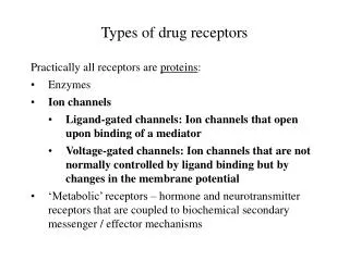
Why Glutamate Receptors are Important in Neurology:. Glutamate is present in millimolar quantities in most cells, including neurons and glia Glutamate is the main excitatory neurotransmitter in the mammalian CNS Glutamate is released in large quantities during Stroke Trauma Epilepsy

E N D
Why Glutamate Receptors are Important in Neurology: • Glutamate is present in millimolar quantities in most cells, including neurons and glia • Glutamate is the main excitatory neurotransmitter in the mammalian CNS • Glutamate is released in large quantities during • Stroke • Trauma • Epilepsy • Possibly in chronic neurological disorders
Why Glutamate Receptors are Important in Neurology: • Excess glutamate is released at the synapse through • Synaptic activity • Reverse operation of glutamate transporters • Reduced re-uptake (due to reduced ATP levels) • Glutamate levels may rise at the synapse to hundreds of micromolar, which is enough to cause excitotoxicity
What happens to neurons with excess glutamate? Normal Neuron
What happens to neurons with excess glutamate? • Cell Swelling • Dendritic Beading • Axons: no change (?) Glutamate
Calcium Homeostasis • Ca ions are ubiquitous intracellular 2nd messengers responsible for a multitude of cellular functions including • Activation of numerous enzymes responsible for • Gene expression • Protein structure • Metabolic functions (libids, carbohydrates) • The control of differentiation, polarity, synaptogenesis • Synaptic efficacy – neuronal function & activity
Calcium Homeostasis • For these reasons cells maintain a very tight control of Ca ions • [Ca2+]I : [Ca2+ ] e is 1 : 20,000 • Ca2+ ions are sequestered into intracellular organelles • Ca2+ ions are actively pumped in and out of cellular compartments • Cells contain diverse Ca2+ buffering molecules to restrict the diffusion of Ca2+ ions.
Calcium Neurotoxicity “Ca2+ Excess” is felt to be deleterious to neurons How much is too much remains controversial It is likely that Ca2+ ions activate distinct 2nd messenger signaling pathways in neurons that cause them to die. Excitotoxicity causes “Ca2+ Excess”.
Hypothetical Scheme Leading to Ca Excess: The Case of Neurons
Scheme Leading to Ca Excess:The case of axons (white matter)
Neurotoxic Phenomena triggered by Calcium Excess • The formation of free radical species • Nitric Oxide formation • Calcium Activated Proteases • Endonucleases, Apoptosis, Necrosis • Mitochondrial Damage • Acidosis
Free radicals • Free radicals are reactive oxygen species having a single unpaired electron: • e.g.: Superoxide (O2-), hydroxyl (OH-) • Free radicals produce damage by reacting (oxidizing) with critical cellular elements, usually structural proteins, membrane lipids, DNA. • Free radicals are produced mostly in mitochondria.
Superoxide production: Although molecular oxygen is reduced to water in the terminal complex IV by a sequential four-electron transfer, a minor proportion can be reduced by a 1e addition that occurs predominantly in complex III but also in complex I. A chance exists that this second electron can be transferred to molecular oxygen, generating the superoxide anion O2·. Thus- normal mitochondria produce a small amount of superoxide. This superoxide is normally scavenged by superoxide dismutase (SOD)
Excitotoxicity and ROS: Calcium loading of isolated mitochondria increases the production of O2· Excototoxicity causes mitochondrial Ca loading.
Nitric Oxide Production NO is a gas with a half-life of 6s. It is produced in: - Vascular endothelium (vasorelaxant) - Glial cells - Neurons It is considered by many to be a neurotransmitter associated with processes related to synaptic plasticity, learning and memory.
NO toxicity: NO is a relatively innocuous gas. However, when combined with superoxide: NO + O2- = ONOO- ONOO- is a highly reactive free radical species that produces damage in neurons.
Calcium activated proteases MAP2 immunofluorescence Controls NMDA Recovery
Calcium activated proteases (Caplains) Role unclear – felt by most to mediate neuronal damage in stroke. However, some research suggests the reverse- that they may be necessary for neuronal recovery from stroke.
Calcium-activated proteases Calcium-dependent proteolysis contributes to recovery of dendritic structure after NMDA exposure. Calpain activation is not necessarily detrimental and may play a role in dendritic remodeling after neuronal injury. No Calpain Inhibitor Calpain Inhibitor
Endonucleases, apoptosis, necrosis Necrosis: Acute cell death characterized by cell & organelle swelling. Is generally rapid, and occurs due to massive insults. Apoptosis: Slower cell death, characterized by cell shrinkage, nuclear fragmentation, and may be mediated by a “death sequence” dictated by a genetic program.
Endonucleases, apoptosis, necrosis Endonucleases are thought to be calcium-activated enzymes that cleave DNA May be responsible in triggering apoptosis.
How to treat stroke? Concept of therapeutic window: Increases with increased flow. Exact time unknown for humans.
How to treat stroke? • Repair the plumbing • Make the tissue more resilient to poor plumbing.
The plumbing: Best treatment of plumbing failure is prevention. • Risk factors for atherosclerosis • Diabetes • High blood pressure • Hypercholesterolemia • Smoking
The plumbing: Benefit of carotid endarterectomy in patients with symptomatic moderate or severe stenosis. North American Symptomatic Carotid Endarterectomy Trial Collaborators. N Engl J Med 1998 Nov 12;339(20):1415-25
Plumbing After Stroke Onset: Tissue plasminogen activator for acute ischemic stroke. The National Institute of Neurological Disorders and Stroke rt-PA Stroke Study Group. N Engl J Med 1995 Dec 14;333(24):1581-7 “treatment with intravenous t-PA within three hours of the onset of ischemic stroke improved clinical outcome at three months.”
Plumbing After Stroke Onset: Intra-arterial pro-urokinase for acute ischemic stroke: The PROACT II Study: A randomized controlled trial. JAMA (282) 21, December 1, 1999